. 2016 Apr 18;3(3-4):122-33.
doi: 10.18632/oncoscience.302. eCollection 2016.

Quantitative ultrasound imaging of therapy response in bladder cancer in vivo

Affiliations

Quantitative ultrasound imaging of therapy response in bladder cancer in vivo

William T Tran et al. Oncoscience. .

Abstract

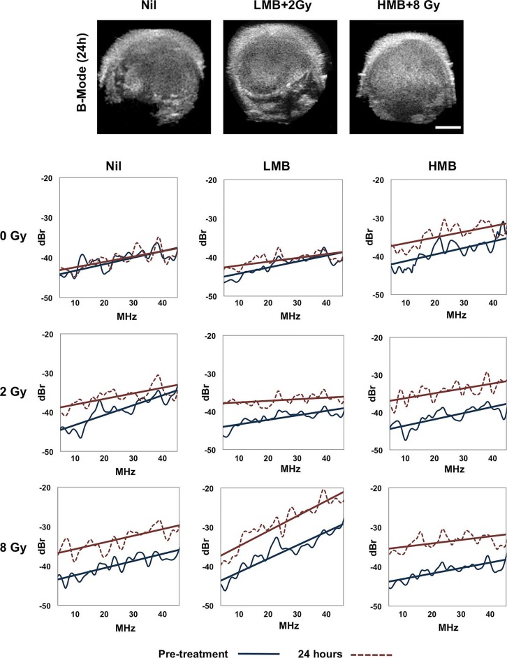
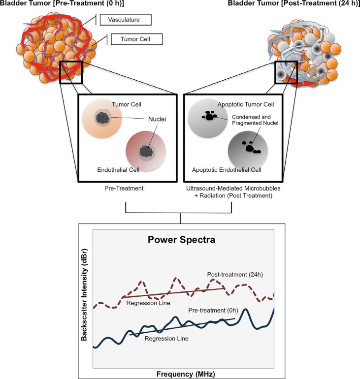
Background and aims: Quantitative ultrasound (QUS) was investigated to monitor bladder cancer treatment response in vivo and to evaluate tumor cell death from combined treatments using ultrasound-stimulated microbubbles and radiation therapy.

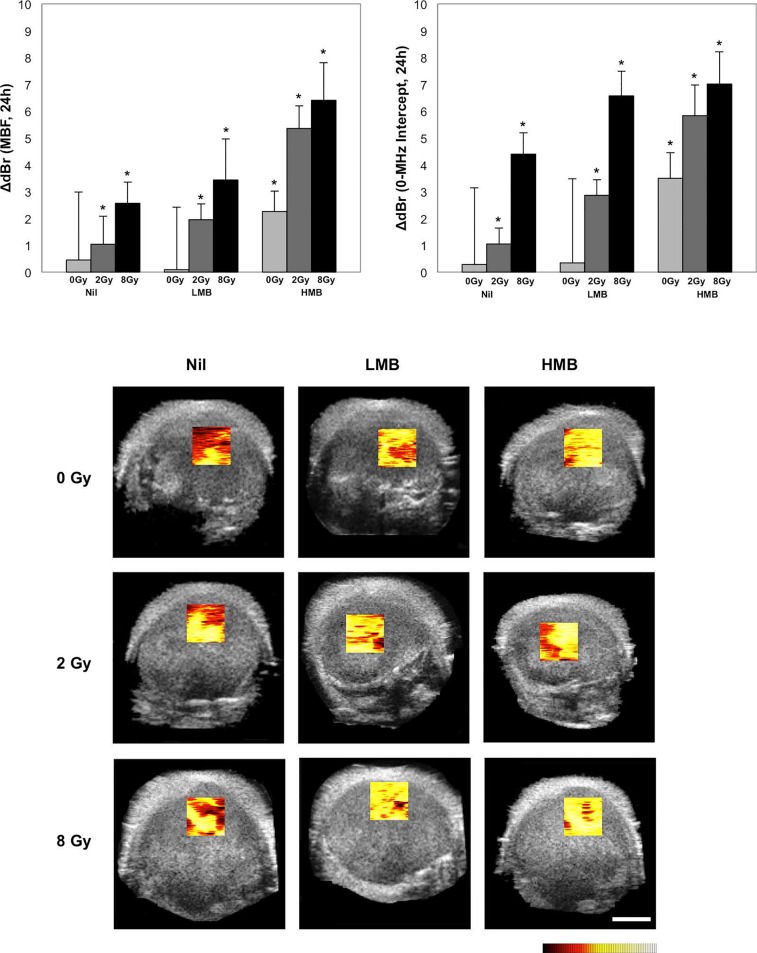
Methods: Tumor-bearing mice (n=45), with bladder cancer xenografts (HT- 1376) were exposed to 9 treatment conditions consisting of variable concentrations of ultrasound-stimulated Definity microbubbles [nil, low (1%), high (3%)], combined with single fractionated doses of radiation (0 Gy, 2 Gy, 8 Gy). High frequency (25 MHz) ultrasound was used to collect the raw radiofrequency (RF) data of the backscatter signal from tumors prior to, and 24 hours after treatment in order to obtain QUS parameters. The calculated QUS spectral parameters included the mid-band fit (MBF), and 0-MHz intercept (SI) using a linear regression analysis of the normalized power spectrum.

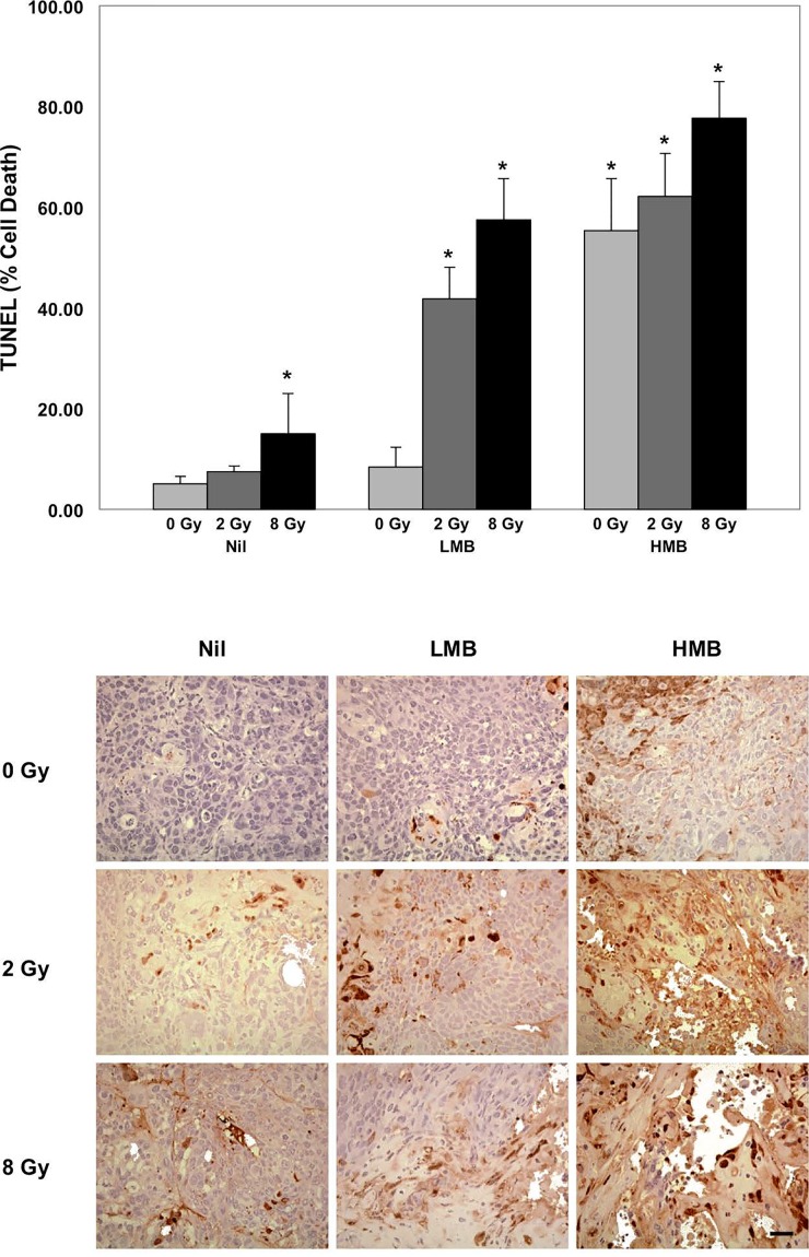
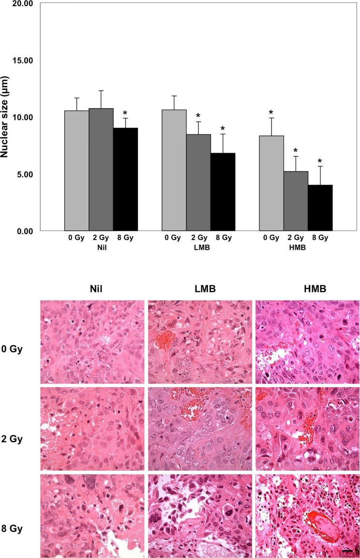
Results and conclusions: There were maximal increases in QUS parameters following treatments with high concentration microbubbles combined with 8 Gy radiation: (ΔMBF = +6.41 ± 1.40 (±SD) dBr and SI= + 7.01 ± 1.20 (±SD) dBr. Histological data revealed increased cell death, and a reduction in nuclear size with treatments, which was mirrored by changes in quantitative ultrasound parameters. QUS demonstrated markers to detect treatment effects in bladder tumors in vivo.

Keywords: quantitative ultrasound; radiation therapy; ultrasound; vascular disrupting agents.


Conflict of interest statement

CONFLICTS OF INTEREST

The authors declare no conflict of interest.

Figures

Figure 1
Figure 1. Representative B-Mode US of tumors and power spectra of samples under treatment conditions
Power spectrum analysis was conducted at baseline (pre-treatment) and 24 hours post treatment. Changes in power spectrum were observed in treatments where higher doses of ultrasound mediated microbubbles and radiation were administered. For spectral parameters, the −6 dB window corresponded to a frequency range approximately 13-35 MHz. Red line = Post-treatment (24h), Blue line = Pre-treatment, US Scale bar = 2mm, Nil= no microbubbles, LMB= Low microbubble concentration (1% v/v), HMB=High microbubble concentration (3% v/v).
Figure 2
Figure 2. Changes in quantitative ultrasound parameters with treatment and corresponding parametric maps of the mid-band fit
Significant increases in the mid-band fit (MBF) and 0-MHz intercept (SI) were observed in higher doses of ultrasound microbubbles and radiation, corresponding to central locations in the tumor. Tumors were treated with single doses of radiation (0 Gy, 2 Gy, 8 Gy) or microbubbles ( Nil, LMB, and HMB) or a combination of both treatment modalities. *p<0.05, when compared to pre-treatment values. Color scale represents a range of 20 dBr. Scale bar = 2 mm. Factorial ANOVA demonstrated significant treatment effects (radiation, microbubbles, radiation*microbubbles) on both the MBF and SI (p<0.001).
Figure 3
Figure 3. High magnification (TUNEL) staining at 24 hours after treatment and quantification of tumor cell death
Treatment effects were observed in combination treatments. Increasing treatment doses resulted in higher areas of tumor cell death. Histology: Top Row. Microbubble treatments alone showed elevated levels of cell death with increased ultrasound-driven microbubbles treatment doses. Middle Row. 2 Gray radiation and Microbubble treatments. Additive effects are observed showing regions increased regions of cell death following higher doses of combination treatment. Bottom Row. Elevated regions of apoptosis as a result of microbubble treatment and radiation. Highest combination doses show areas of cell death and tumor cell death. Magnification=40x, Bar= 25 μm; *=p<0.05, compared to treatment controls (Nil + 0 Gy).
Figure 4
Figure 4. Hematoxylin and eosin staining, and nuclear size assessment
Nuclear size was assessed in tumor cross-sections following 24 hours of treatment. Combination treatments demonstrated a significant difference in nuclear size compared to control samples (Nil + 0 Gy). Histology: Treatment from ultrasound-mediated microbubbles demonstrated vascular disruption, marked by areas with erythrocytes. Aggressive treatment doses showed a decrease in cellularity. Magnification=40x, Bar= 25 μm; *=p<0.05, compared to treatment controls (Nil + 0 Gy).
Figure 5
Figure 5. Schematic representation of QUS analysis in bladder cancer xenografts
Bladder tumors were imaged prior to, and after 24 hours of treatment with ultrasound mediated microbubbles and radiation. Vascular disruption from microbubble cavitation resulted in endothelial cell death (apoptosis) followed by radiation-induced cell death in tumor cells. Spectrum analysis of tumors after 24 hours demonstrate an increase in the backscatter intensity likely caused by fragmented and condensed nuclear structures from both tumor cells and endothelial cells.

Similar articles

Cited by

References

    1. James ND, Hussain SA, Hall E, Jenkins P, Tremlett J, Rawlings C, Crundwell M, Sizer B, Sreenivasan T, Hendron C, Lewis R, Waters R, Huddart RA, Investigators BC. Radiotherapy with or without chemotherapy in muscle-invasive bladder cancer. N Engl J Med. 2012;366:1477–1488. - PubMed
    1. Stenzl A, Cowan NC, De Santis M, Kuczyk MA, Merseburger AS, Ribal MJ, Sherif A, Witjes JA, European Association of U Treatment of muscle-invasive and metastatic bladder cancer: update of the EAU guidelines. Eur Urol. 2011;59:1009–1018. - PubMed
    1. Pectasides D, Pectasides M, Nikolaou M. Adjuvant and neoadjuvant chemotherapy in muscle invasive bladder cancer: literature review. Eur Urol. 2005;48:60–67. discussion 67-68. - PubMed
    1. Wilson WR, Li AE, Cowan DS, Siim BG. Enhancement of tumor radiation response by the antivascular agent 5,6-dimethylxanthenone-4-acetic acid. Int J Radiat Oncol Biol Phys. 1998;42:905–908. - PubMed
    1. Tran WT, Iradji S, Sofroni E, Giles A, Eddy D, Czarnota GJ. Microbubble and ultrasound radioenhancement of bladder cancer. British journal of cancer. 2012;107:469–476. - PMC - PubMed