Review
. 2016 May 26;19(6):771-83.
doi: 10.1038/nn.4288.

Pericytes of the neurovascular unit: key functions and signaling pathways

Affiliations
Review

Pericytes of the neurovascular unit: key functions and signaling pathways

Melanie D Sweeney et al. Nat Neurosci. .

Abstract

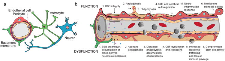
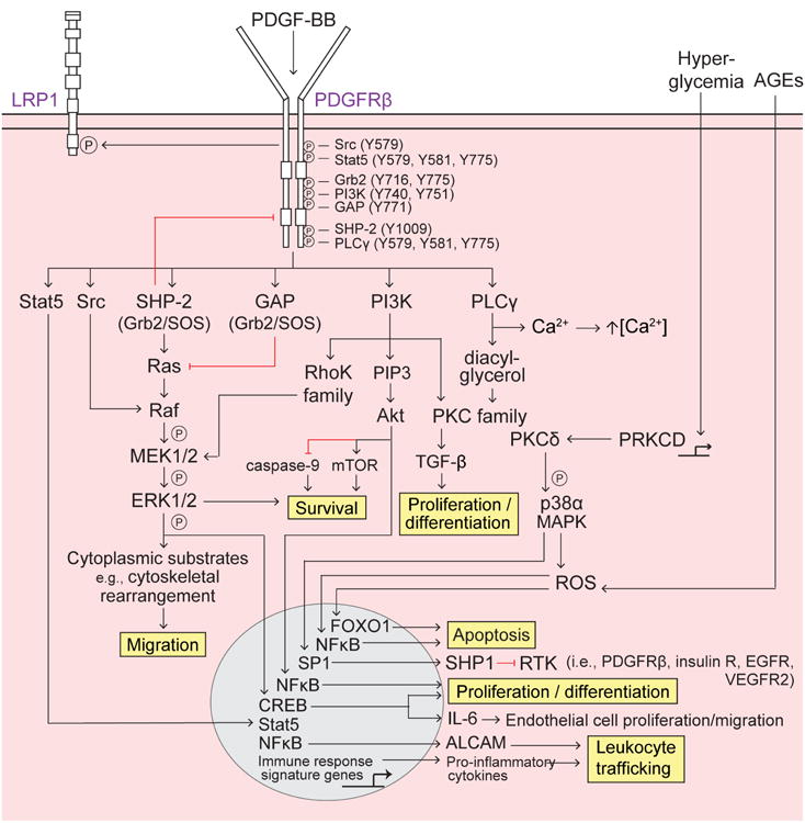
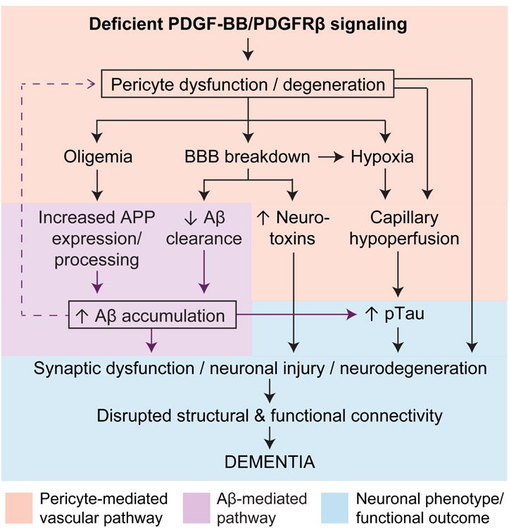
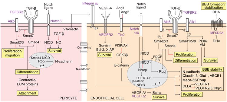
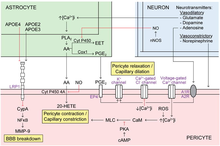
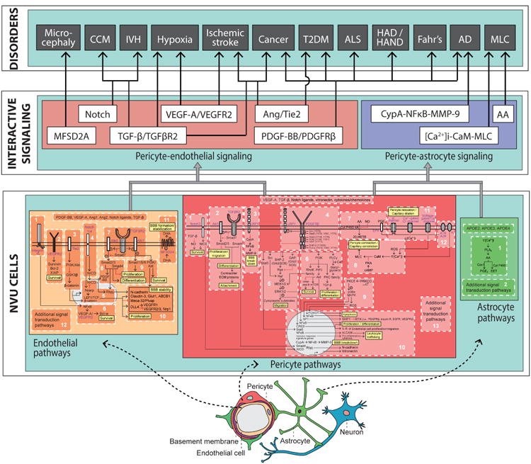
Pericytes are vascular mural cells embedded in the basement membrane of blood microvessels. They extend their processes along capillaries, pre-capillary arterioles and post-capillary venules. CNS pericytes are uniquely positioned in the neurovascular unit between endothelial cells, astrocytes and neurons. They integrate, coordinate and process signals from their neighboring cells to generate diverse functional responses that are critical for CNS functions in health and disease, including regulation of the blood-brain barrier permeability, angiogenesis, clearance of toxic metabolites, capillary hemodynamic responses, neuroinflammation and stem cell activity. Here we examine the key signaling pathways between pericytes and their neighboring endothelial cells, astrocytes and neurons that control neurovascular functions. We also review the role of pericytes in CNS disorders including rare monogenic diseases and complex neurological disorders such as Alzheimer's disease and brain tumors. Finally, we discuss directions for future studies.


Conflict of interest statement

Notes: The authors declare no conflict of interest.

Figures

Figure 1
Figure 1. The multi-functional role of CNS pericytes at the neurovascular unit (NVU)
(a) A simplified NVU diagram showing the interactive cellular network at the level of brain capillaries that comprises vascular cells (e.g., pericytes and endothelial cells), glial cells (e.g., astrocytes), and neurons. Intricate cell-cell communication and signal transduction mechanisms of NVU cell types are highly controlled to regulate numerous functions in the CNS. (b) Under physiological conditions (top row), pericytes regulate 1) blood-brain barrier (BBB) integrity, i.e., tight/adherens junctions and transcytosis across the BBB; 2) angiogenesis, i.e. microvascular remodeling, stability, and architecture; 3) phagocytosis, i.e., clearance of toxic metabolites from the CNS; 4) cerebral blood flow (CBF) and capillary diameter; 5) neuroinflammation, i.e., leukocyte trafficking into the brain; and 6) multipotent stem cell activity. Pericyte dysfunction (bottom row) is characterized by 1) BBB breakdown causing leakage of neurotoxic blood-derived molecules into the brain (e.g., fibrinogen, thrombin, plasminogen, erythrocyte-derived free iron, and anti-brain antibodies); 2) aberrant angiogenesis; 3) impaired phagocytosis causing CNS accumulation of neurotoxins; 4) CBF dysfunction and ischemic capillary obstruction; 5) increased leukocyte trafficking promoting neuroinflammation; and 6) impaired stem cell-like ability to differentiate into neuronal and hematopoietic cells. Pericyte dysfunction is present in numerous neurological conditions and can contribute to the disease pathogenesis.
Figure 2
Figure 2. PDGF-BB/PDGFRp signaling in pericytes
Platelet-derived growth factor-BB (PDGF-BB) secreted by endothelial cells binds to the PDGF receptor-β (PDGFRβ) on pericytes, causing receptor dimerization, autophosphorylation, and activation. Several Src homology 2 (SH2) domain-containing proteins (Src, Stat5, Grb2, phosphatidyl inositol 3-phosphate (PI3K), GTPase activating protein (GAP), SH2 tyrosine phosphatase (SHP-2), and phospholipase Cγ (PLCγ) bind to distinct phosphorylated (P) tyrosine (Y) residues SH2 domain-containing proteins bound to PDGFRβ differentially activate downstream signaling pathways to regulate pericyte survival, migration, apoptosis, proliferation and differentiation, as well as leukocyte trafficking, described as follows: Survival – promoted via PI3K-Akt activation of mammalian target of rapamycin (mTOR) and inhibition of caspase-9 and the SHP-2-mediated MAPK pathway; Migration – SHP-2-mediated MAPK pathway promotes cytoskeletal rearrangement and cell migration. Src activated Raf synergistically activates the MAPK pathway whereas GAP inhibition of Ras decreases MAPK signaling; Apoptosis – Extracellular advanced glycation endproducts (AGEs) induce intracellular reactive oxygen species (ROS) and FOXO1-mediated apoptosis, and PRKCD transcriptional expression of protein kinase C-δ (PKCδ) activates p38α MAPK to induce downstream production of ROS and mitochondrial cytochrome c release, resulting in apoptosis; Proliferation and differentiation – promoted by the PI3K pathway, specifically via PKC-TGF-β and PIP3-Akt transcriptional activation of NFκB; Leukocyte trafficking – PDGFRβ regulates pro-inflammatory responses, e.g., peripheral leukocyte trafficking into the CNS, via transcriptional expression of immune response signaling genes (e.g., cytokines and chemokines) and also via Akt-induced activation of NFκB and transcriptional expression of the novel activated leukocyte cell adhesion molecule (ALCAM).
Figure 3
Figure 3. Deficient PDGF-BB/PDGFRp signaling in pericytes: A possible role in promoting neuronal dysfunction and degeneration in Alzheimer's dementia
In the amyloid-β ()-independent pathway (pink box), a deficient PDGF-BB/PDGFRβ signaling leads to pericyte dysfunction and/or degeneration resulting in microvascular and cerebral blood flow (CBF) reductions and oligemia (brain hypoperfusion), from one hand, and blood-brain barrier (BBB) breakdown with accumulation of blood-derived toxic products in the brain, from the other. BBB breakdown leads to capillary edema contributing to capillary hypoperfusion and hypoxia. In the Aβ- pathway (purple box), oligemia leads to increased Aβ production, whereas BBB breakdown and deficient PDGFRβ signaling can both lead to faulty Aβ clearance, which in turn promotes Aβ accumulation in the brain. Synergistic action of Aβ-independent and Aβ pathway lead to accelerated tau hyperphosphorylation, formation of neurofibrillary tangles, synaptic dysfunction and loss, and neuronal degeneration, which altogether promotes behavioral deficits and dementia (blue box).
Figure 4
Figure 4. TGF-β/TGFβR2, Notch, VEGF-A/VEGFR2, Ang/Tie2 and MFSD2A signaling pathways
Pericytes (left, pink) – Tumor growth factor-β (TGF-β) secreted by endothelial cells and pericytes binds to TGF-β receptor 2 (TGFβR2) that phosphorylates activin-like kinase 5 (Alk5) (inhibited by Smad7) activating the downstream Smad signaling cascade. Activated Smad2/3 inhibits pericyte proliferation and migration. Recruitment of Smad4 to Smad2/3 complex transcriptionally promotes pericyte differentiation, expression of contractile/extracellular matrix (ECM) proteins, and pericyte attachment. TGF-β also inhibits nitric oxide (NO) generation promoting survival. Activated Notch3 receptor cleaves its Notch intracellular domain (NICD), which promotes survival. Notch-NICD pathway works cooperatively with TGF-β/TGFβR2-Smad-4 pathway to stimulate Rbpj-mediated expression of N-cadherin that increases blood-brain barrier (BBB) stability. Endothelial cells (right, orange) – VEGF-A/VEGFR2 autocrine/paracrine pathway promotes survival via increased expression of anti-apoptotic Bcl-2, Survivin, and X-linked inhibitor of apoptosis protein (XIAP). Vitronectin secreted by pericytes acts on integrin aV on endothelial cells resulting in NFκB-mediated transcriptional expression of VEGF-A and intracrine-mediated VEGF-A/VEGFR2-dependent Bcl-w expression promoting survival. Pericyte-derived Ang1 acts on endothelial Tie2 receptor, and endothelial-secreted Ang2 blocks Ang1 binding to Tie2, acting as a Tie2 antagonist. Ang1/Tie2 activates phosphatidyl inositol 3-phosphate (PI3K)/Akt pathway resulting in inhibition of glycogen synthase kinase 3β (GSK3P), an inhibitor of β-catenin; this leads to β-catenin nuclear translocation resulting in activation of TCF/LEF transcription factors that control expression of several proteins promoting BBB stability (shown in a box). Notch1/4 contributes to β-catenin-mediated BBB stability via the Notch-regulated ankyrin repeat protein (Nrarp), which increases β-catenin nuclear signaling by inhibiting LEF1 degradation and decreases Notch signaling via NICD destabilization. Additionally, Notch1/4-NICD pathway stimulates PDGF-BB expression and RBP-Jκ-mediated expression of N-cadherin contributing to BBB stability. As in pericytes, TGF-β/TGFβR2 pathway in endothelium similarly activates i) Alk5-Smad2/3/4 complex to transcriptionally promote differentiation, inhibit proliferation, and induce RBP-Jκ-mediated expression of N-cadherin, ii) Alk1-Smad1/5/8 to promote proliferation, and iii) Alk1-PI3K/Akt pathway to promote survival and BBB stability. The major facilitator superfamily domain-containing protein 2a (MFSD2A) facilitates luminal-to-abluminal transport of docosahexaenoic acid (DHA), an essential omega-3 fatty acid, and controls formation of the BBB; its expression depends on the presence of pericytes.
Figure 5
Figure 5. Pericyte-astrocyte and pericyte-neuron signaling pathways
Astrocytes (top left, green) secrete apolipoprotein E (APOE) 2 and 3 that bind to pericyte lipoprotein LRP1 receptor (bottom, orange) to inhibit downstream CypA-NFκB-MMP-9 pathway. In contrast, APOE4 binds weakly to LRP1, which activates the pro-inflammatory CypA-NFκB-MMP-9 cascade leading to blood-brain barrier (BBB) breakdown. Astrocyte intracellular Ca2+ concentration ([Ca2+]i) increases in response to neuronal factors, for example glutamate, which promotes phospholipase A2 (PLA2)-mediated arachidonic acid (AA) generation. In astrocytes, AA is metabolized into prostaglandin E2 PGE2) via cyclooxtgenase-1 (Cox1), as well as into epoxyeicosotrienoic acids (EET) via cytochrome P450. Astrocytic AA is metabolized into 20-HETE in mural cells via membrane-bound cytochrome P450 4A, which promotes pericyte contraction. PGE2 from astrocytes binds to pericyte EP4 receptor, which alters K+ conductance and promotes pericyte relaxation. Nitric oxide (NO) generated by neurons inhibits cytochrome P450 in astrocytes and cytochrome P450 4A in pericytes to prevent AA to EET and AA to 20-HETE metabolism, respectively. In pericytes, increased cyclic adenosine monophosphate (cAMP) signals via protein kinase A (PKA) to inhibit myosin light chain (MLC) phosphorylation and prevent pericyte contraction. Additionally, pericyte [Ca2+]i increases in response to voltage-gated Ca2+ channels and reactive oxygen species (ROS). Increased [Ca2+]i in pericytes is shown to promote contraction, possibly via downstream signaling through calmodulin (CaM) and MLC kinase (MLCK) which phosphorylates MLC to induce contraction, as shown in VSMCs. Conversely, decreasing [Ca2+]i in pericytes inhibits Ca2+-gated Cl- channels which promotes relaxation. Furthermore, neurotransmitters promote pericyte, relaxation (e.g., glutamate, dopamine, and adenosine) or contraction (e.g., norepinephrine). For example, adenosine signals through adenosine A1 and A2 receptors (A1R, A2R) on pericytes to alter K+ conductance and promote pericyte relaxation.
Figure 6
Figure 6. Integrated pathways between pericytes, endothelial cells and astrocytes within the neurovascular unit (NVU)
A proposed three-layered model of the NVU. The first layer, ‘NVU cells,’ is the foundational layer of cell type-specific systems, each of which consists of integrated modular molecular pathways. Here, we show the endothelial cell (yellow) partitioned into 11 pathways, the pericyte (pink) partitioned into 12 pathways, and the astrocyte (green) partitioned into 1 pathway. Each major signal transduction pathway within each NVU cell type also provides for the modular addition of new signaling pathways, denoted as “additional signal transduction pathways.” The second layer, ‘Interactive Signaling,’ instantiates the converging points of interactions of key signaling pathways between pericytes-endothelial cells and pericytes-astrocytes. The pericyte-endothelial signaling (coral box) at the second layer consists of 6 major signaling pathways: MFSD2A, Notch, TGF-β/TGFβR2, VEGF-A/VEGFR2, Ang/Tie2, and PDGF-BB/PDGFRβ. The pericyte-astrocyte signaling (purple box), also at the second layer, consists of 3 major signaling pathway: CypA-NFκB-MMP-9, arachidonic acid (AA), and [Ca2+]i-CaM-MLC. The third layer, ‘Disorders,’ proposes major signaling pathways of CNS pericytes with neighboring NVU cell types (i.e., endothelial cells and astrocytes) that are suggested to contribute to pericytes dysfunction in neurological disorders, including: Microcephaly, cerebral cavernous malformation (CCM), intraventricular hemorrhage (IVH), hypoxia, ischemic stroke, cancer, type 2 diabetes mellitus (T2DM), amyotrophic lateral sclerosis (ALS), HIV-associated dementia (HAD) and HIV-associated neurocognitive disorders (HAND), Fahr's disease, Alzheimer's disease (AD), and megalencephalic leukoencephalopathy with subcortical cysts (MLC).

References

    1. Zlokovic BV. Neurovascular pathways to neurodegeneration in Alzheimer's disease and other disorders. Nat Rev Neurosci. 2011;12:723–738. - PMC - PubMed
    1. Winkler EA, Bell RD, Zlokovic BV. Central nervous system pericytes in health and disease. Nat Neurosci. 2011;14:1398–1405. - PMC - PubMed
    1. Iadecola C. The pathobiology of vascular dementia. Neuron. 2013;80:844–866. - PMC - PubMed
    1. Attwell D, et al. Glial and neuronal control of brain blood flow. Nature. 2010;468:232–243. - PMC - PubMed
    1. Armulik A, Genové G, Betsholtz C. Pericytes: developmental, physiological, and pathological perspectives, problems, and promises. Dev Cell. 2011;21:193–215. - PubMed

Publication types