

Influence of Omega 3 and 6 Fatty Acids on Human Meibomian Gland Epithelial Cells
- PMID: 27227397
- PMCID: PMC4938711
- DOI: 10.1097/ICO.0000000000000874
Influence of Omega 3 and 6 Fatty Acids on Human Meibomian Gland Epithelial Cells
Abstract
Purpose: Oral supplementation with omega 3 (ω-3) and/or 6 (ω-6) fatty acids (FAs) has been reported to alleviate the signs and symptoms of dry eye disease and to improve the expressibility and quality of meibum, in patients with meibomian gland dysfunction. We tested our hypothesis that these FA effects may reflect a direct FA action on human meibomian gland epithelial cells.
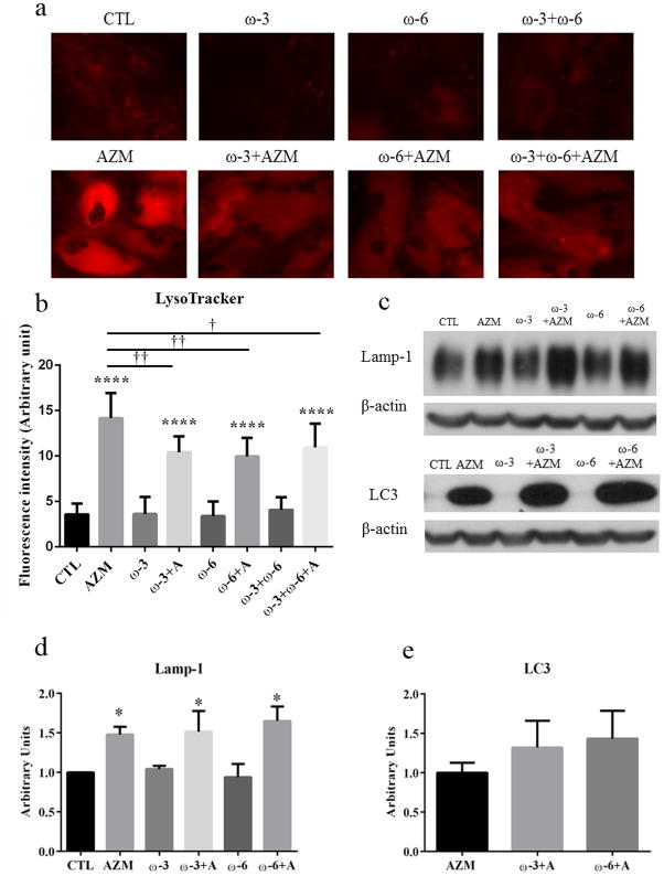
Methods: Immortalized human meibomian gland epithelial cells (IHMGECs) were cultured with ω-3, ω-6, or both FAs together for up to 7 days in the presence or absence of serum. After FA exposure, cells were analyzed for lipid expression, lysosome content, and proliferative ability.
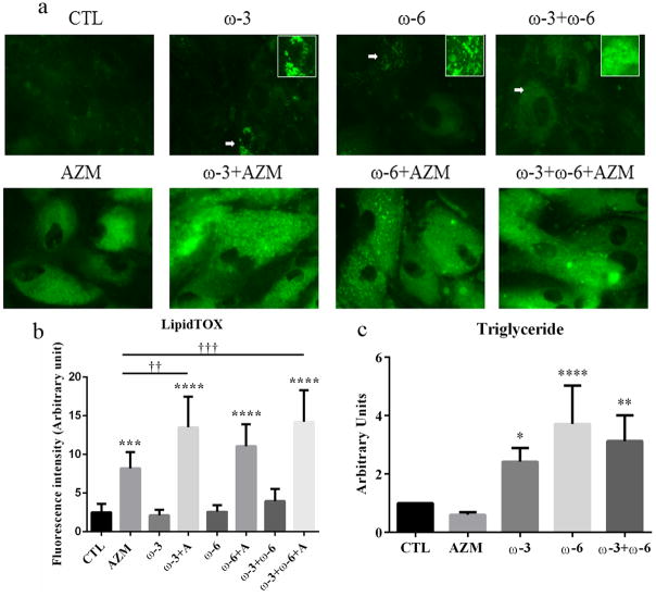
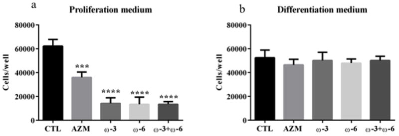
Results: Our research shows that ω-3 and ω-6 stimulate the accumulation of small neutral lipid-containing vesicles, but not lysosomes, in IHMGECs. This vesicular effect was associated with a 2.4- to 3.7-fold increase in the cellular content of triglycerides after ω-3 and ω-6 treatment, respectively. The combination of both FAs together also enhanced triglyceride levels. Of interest, culture of IHMGECs with ω-3 and azithromycin, a known inducer of IHMGEC differentiation, led to a significantly greater amount of total neutral lipids, relative to that found with azithromycin alone. Cellular exposure to the FAs did not alter the expression of free or esterified cholesterol, or phospholipids. Further, these FAs, alone or together, prevented the proliferation of IHMGECs in serum-free, but not serum-containing, media.
Conclusions: Our findings support our hypothesis and demonstrate that ω-3 and ω-6 can act directly on IHMGECs to influence the quality and quantity of intracellular lipids.
Conflict of interest statement
A patent application has been filed related to azithromycin. The intellectual property for this application is owned by the Schepens Eye Research Institute/Massachusetts Eye and Ear. Otherwise, the authors have no conflict of interest.
Figures
References
-
- The definition and classification of dry eye disease: report of the Definition and Classification Subcommittee of the International Dry Eye WorkShop (2007) Ocul Surf. 2007;5:75–92. - PubMed
-
- Chao W, Belmonte C, Benitez Del Castillo J, et al. Report of the Inaugural Meeting of the TFOS i2 = initiating innovation Series: Targeting the Unmet Need for Dry Eye Treatment (London, United Kingdom, March 21, 2015) Ocul Surf. 2016 - PubMed
-
- Lemp MA, Crews LA, Bron AJ, et al. Distribution of aqueous-deficient and evaporative dry eye in a clinic-based patient cohort: a retrospective study. Cornea. 2012;31:472–478. - PubMed
MeSH terms
Substances
Grants and funding
LinkOut - more resources
Full Text Sources
Other Literature Sources
Research Materials
Miscellaneous
