. 2016 May 26;11(5):e0156218.
doi: 10.1371/journal.pone.0156218. eCollection 2016.

Crystal Structure of the FERM-SH2 Module of Human Jak2

Affiliations

Crystal Structure of the FERM-SH2 Module of Human Jak2

Randall McNally et al. PLoS One. .

Abstract

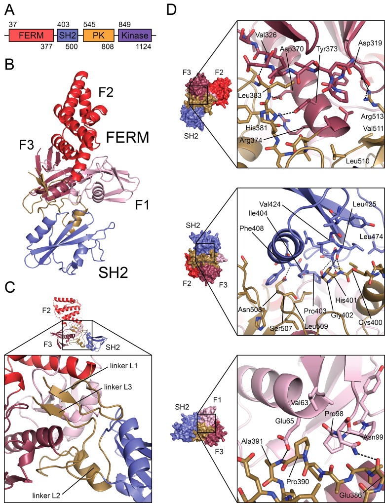
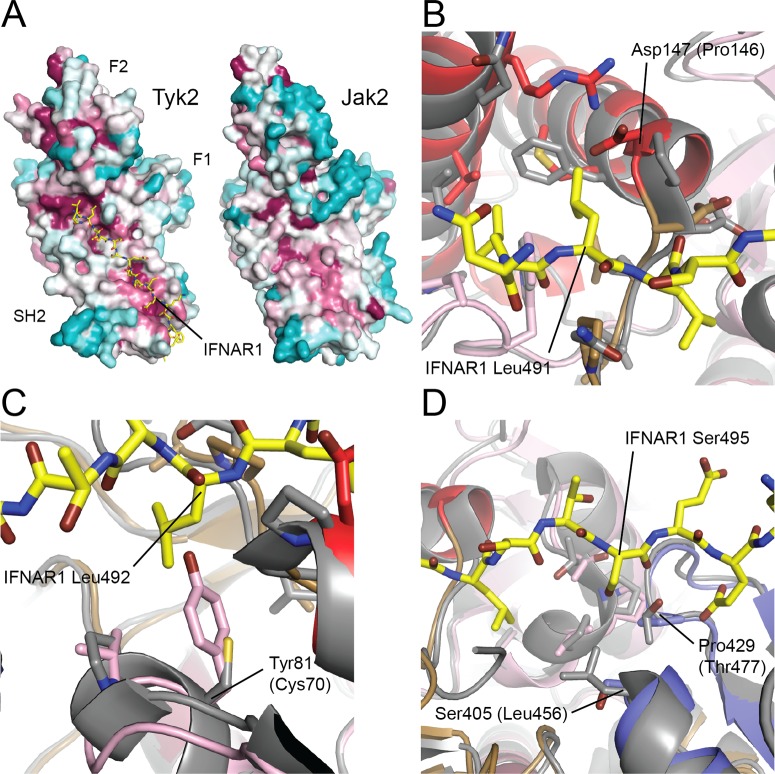
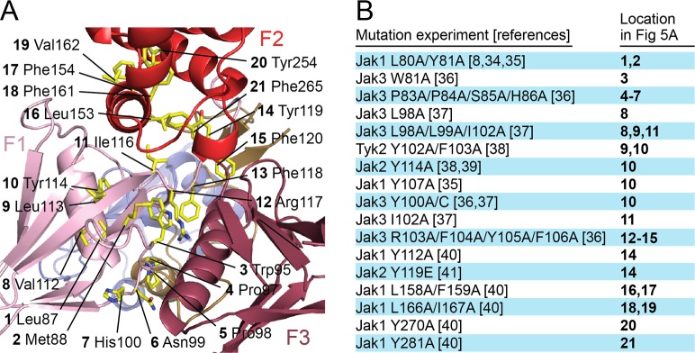
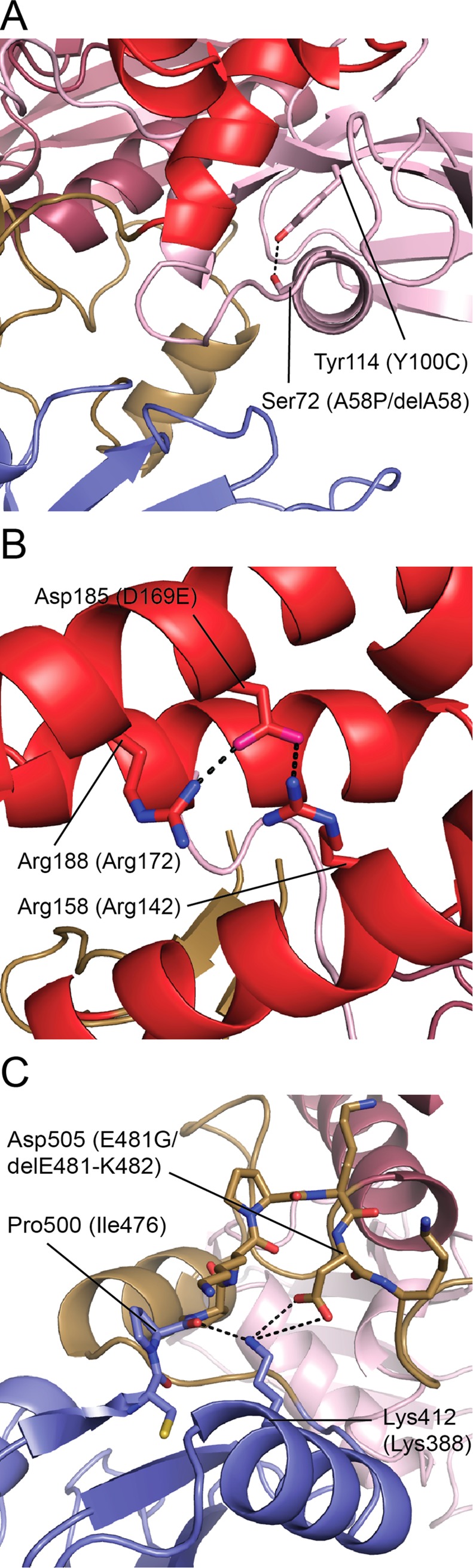
Jak-family tyrosine kinases mediate signaling from diverse cytokine receptors. Binding of Jaks to their cognate receptors is mediated by their N-terminal region, which contains FERM and SH2 domains. Here we describe the crystal structure of the FERM-SH2 region of Jak2 at 3.0Å resolution. The structure reveals that these domains and their flanking linker segments interact intimately to form an integrated structural module. The Jak2 FERM-SH2 structure closely resembles that recently described for Tyk2, another member of the Jak family. While the overall architecture and interdomain orientations are preserved between Jak2 and Tyk2, we identify residues in the putative receptor-binding groove that differ between the two and may contribute to the specificity of receptor recognition. Analysis of Jak mutations that are reported to disrupt receptor binding reveals that they lie in the hydrophobic core of the FERM domain, and are thus expected to compromise the structural integrity of the FERM-SH2 unit. Similarly, analysis of mutations in Jak3 that are associated with severe combined immunodeficiency suggests that they compromise Jak3 function by destabilizing the FERM-SH2 structure.


Conflict of interest statement

Competing Interests: The authors have declared that no competing interests exist.

Figures

Fig 1
Fig 1. Crystal structure of the FERM-SH2 domains of Jak2.
(A) The domain structure of Jak-family kinases; residue numbers correspond to human Jak2. ‘SH2’, SH2-like. (B) Structure of Jak2 FERM-SH2 domains, with domains and sub-domains indicated. (C) Detail of Jak-specific insertions, linkers L1, L2, and L3. L1 connects FERM F1 and F2 lobes; linker L2 connects FERM F3 lobe and SH2 domain; linker L3 connects SH2 and pseudokinase domains. (D) Interfaces between Jak-specific insertions and FERM, SH2 domains. Top panel: FERM F3 lobe (dark red) and linkers L2 and L3 (gold). Center panel: SH2 domain (blue) and linkers L2 and L3. Bottom panel: FERM F1 lobe (pink) and linker L2. Dashed lines indicate hydrogen bonds and salt bridges across interfaces. Crystal structure figures were generated using PyMOL.
Fig 2
Fig 2. Comparison of the structures of Jak2 and Tyk2-IFNAR1.
(A) Plot of the displacement of structurally equivalent Cα atoms between superposed structures of Jak2 and Tyk2. (B) Superposition of Jak2 (colored as in Fig 1) and Tyk2-IFNAR1 (Tyk2 is colored gray, IFNAR1 is yellow). (C-E) Detailed views of superposition in (B). (C) The FERM F2 lobe of Jak2 differs from that of Tyk2. The linker between helices F2-α1 and F2-α2 is unstructured in Jak2, while in Tyk2 it forms a 310 helix and N-terminal extension of helix F2-α2. In addition, helices F2-α2’, F2-α2”, and F2-α3 of the Jak2 F2 lobe are displaced relative to Tyk2. (D-E) The FERM F3 lobe of Jak2 differs from that of Tyk2. A C-terminal extension of strand F3-β1 and subsequent linker that forms further interactions with the F3 lobe in Tyk2 are absent in the Jak2 structure. Dashed lines indicate hydrogen bonds and salt bridges. Jak2 residues are labeled with corresponding Tyk2 residues in parentheses.
Fig 3
Fig 3. Insights into the specificity of receptor binding to Jak2 vs. Tyk2.
(A) Sequence conservation among Jak family members across phylogeny, mapped onto the surfaces of Tyk2 and Jak2 FERM-SH2. The surface is shaded from magenta (most conserved) to teal (most variable) on the basis of ConSurf analysis. The surface of Jak2 that corresponds to the highly conserved IFNAR1 binding pocket on Tyk2 is also highly conserved, suggesting that the receptor-binding function of this surface is retained for Jak2. (B-D) Superposition of the structures of Jak2 (colored as in Fig 1) and Tyk2-IFNAR1 (Tyk2 is colored gray, IFNAR1 is yellow) reveal differences that may determine the specificity of receptors that Jak2 and Tyk2 are capable of binding. (B) In the IFNAR1 Leu491 binding pocket of Tyk2, Pro146 is replaced by Asp in Jak2, a charged residue that is incompatible with the hydrophobic IFNAR1 Leu491. (C) In the IFNAR1 Leu492 binding pocket of Tyk2, replacement of Tyk2 Cys70 with Tyr (as in Jak2) results in steric occlusion of IFNAR1 Leu492. (D) In the IFNAR1 Ser495 binding pocket of Tyk2, the polar Tyk2 Thr477 is replaced in Jak2 by the hydrophobic Pro429, while in the base of the pocket the bulky, hydrophobic Tyk2 Leu456 is replaced by the relatively small, polar Ser405 in Jak2.
Fig 4
Fig 4. The Jak2 SH2 domain lacks a typical phosphotyrosine-binding pocket.
The crystal structure of the SH2 domain of the Src-family kinase Lck bound to a high-affinity phosphotyrosyl peptide (PDB ID 1LCJ) [33] is superimposed on the Jak2 SH2 domain. The Lck structure is shown as a white ribbon, with the bound phosphopeptide in green. The Jak2 SH2 domain is colored blue. The phosphotyrosine sidechain and selected residues in the binding pocket are shown in stick form. Note that Phe436 in the Jak2 SH2 domain blocks the position that would be occupied by the phenyl group of a bound phosphotyrosine. Labels refer to Jak2 residues.
Fig 5
Fig 5. Functional mutations in Jak mapped onto the structure of Jak2 FERM-SH2.
(A) Residues in the Jak FERM domain that, when mutated, have been found experimentally to result in deficient binding to cytokine receptor are mapped to their corresponding residues on Jak2 and colored yellow. These residues are largely buried, indicating that the effect on receptor binding from mutating them is likely a result of destabilization of the FERM domain. Labels indicate Jak2 equivalents of mutated residues. (B) List of mutation experiments referred to in (A). Not pictured: SCID mutants Jak3 delA58 (equivalent to Jak2 Ser72), Jak3 D169E (equivalent to Jak2 Asp185), and Jak3 R402H (equivalent to Jak2 Arg426) [36,42].
Fig 6
Fig 6. Jak3 SCID mutations mapped onto structure of Jak2.
(A) Jak2 residues Tyr114 and Ser72, equivalent in position to SCID mutations Jak3 Y100C and A58P, respectively, are buried in the FERM F1 lobe. These mutations, along with SCID mutation Jak3 delA58, likely destabilize the F1 lobe. (B) Jak2 Asp185 makes salt bridge interactions with Arg158 and Arg188. The SCID mutation Jak3 D169E (equivalent in position to Jak2 Asp185) potentially disrupts this structural network. (C) The Jak2 residue Asp505, equivalent in position to SCID mutation Jak3 E481G, participates in a structural network across the L3-SH2 interface with Lys412 and the backbone carbonyl of Pro500. This mutation, as well as SCID mutation delE481-K482, disturbs this network. Dashed lines indicate hydrogen bonds and salt bridges. Jak2 residues are labeled with corresponding Jak3 residues in parentheses.

Similar articles

Cited by

References

    1. Baker SJ, Rane SG, Reddy EP (2007) Hematopoietic cytokine receptor signaling. Oncogene 26: 6724–6737. - PubMed
    1. Stark GR, Darnell JE Jr. (2012) The JAK-STAT pathway at twenty. Immunity 36: 503–514. 10.1016/j.immuni.2012.03.013 - DOI - PMC - PubMed
    1. Ghoreschi K, Laurence A, O'Shea JJ (2009) Janus kinases in immune cell signaling. Immunol Rev 228: 273–287. 10.1111/j.1600-065X.2008.00754.x - DOI - PMC - PubMed
    1. Haan C, Kreis S, Margue C, Behrmann I (2006) Jaks and cytokine receptors—an intimate relationship. Biochem Pharmacol 72: 1538–1546. - PubMed
    1. Murakami M, Narazaki M, Hibi M, Yawata H, Yasukawa K, Hamaguchi M, et al. (1991) Critical cytoplasmic region of the interleukin 6 signal transducer gp130 is conserved in the cytokine receptor family. Proc Natl Acad Sci U S A 88: 11349–11353. - PMC - PubMed

LinkOut - more resources