Complexities in Isolation and Purification of Multiple Viruses from Mixed Viral Infections: Viral Interference, Persistence and Exclusion
- PMID: 27227480
- PMCID: PMC4881941
- DOI: 10.1371/journal.pone.0156110
Complexities in Isolation and Purification of Multiple Viruses from Mixed Viral Infections: Viral Interference, Persistence and Exclusion
Abstract
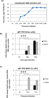
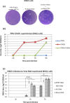
Successful purification of multiple viruses from mixed infections remains a challenge. In this study, we investigated peste des petits ruminants virus (PPRV) and foot-and-mouth disease virus (FMDV) mixed infection in goats. Rather than in a single cell type, cytopathic effect (CPE) of the virus was observed in cocultured Vero/BHK-21 cells at 6th blind passage (BP). PPRV, but not FMDV could be purified from the virus mixture by plaque assay. Viral RNA (mixture) transfection in BHK-21 cells produced FMDV but not PPRV virions, a strategy which we have successfully employed for the first time to eliminate the negative-stranded RNA virus from the virus mixture. FMDV phenotypes, such as replication competent but noncytolytic, cytolytic but defective in plaque formation and, cytolytic but defective in both plaque formation and standard FMDV genome were observed respectively, at passage level BP8, BP15 and BP19 and hence complicated virus isolation in the cell culture system. Mixed infection was not found to induce any significant antigenic and genetic diversity in both PPRV and FMDV. Further, we for the first time demonstrated the viral interference between PPRV and FMDV. Prior transfection of PPRV RNA, but not Newcastle disease virus (NDV) and rotavirus RNA resulted in reduced FMDV replication in BHK-21 cells suggesting that the PPRV RNA-induced interference was specifically directed against FMDV. On long-term coinfection of some acute pathogenic viruses (all possible combinations of PPRV, FMDV, NDV and buffalopox virus) in Vero cells, in most cases, one of the coinfecting viruses was excluded at passage level 5 suggesting that the long-term coinfection may modify viral persistence. To the best of our knowledge, this is the first documented evidence describing a natural mixed infection of FMDV and PPRV. The study not only provides simple and reliable methodologies for isolation and purification of two epidemiologically and economically important groups of viruses, but could also help in establishing better guidelines for trading animals that could transmit further infections and epidemics in disease free nations.
Conflict of interest statement
Figures


Similar articles
-
Isolation, identification and characterization of a Peste des Petits Ruminants virus from an outbreak in Nanakpur, India.J Virol Methods. 2013 May;189(2):388-92. doi: 10.1016/j.jviromet.2013.03.002. Epub 2013 Mar 15. J Virol Methods. 2013. PMID: 23500799
-
Induction of protective immune response against both PPRV and FMDV by a novel recombinant PPRV expressing FMDV VP1.Vet Res. 2014 Jun 4;45(1):62. doi: 10.1186/1297-9716-45-62. Vet Res. 2014. PMID: 24898430 Free PMC article.
-
Development of real-time and lateral flow strip reverse transcription recombinase polymerase Amplification assays for rapid detection of peste des petits ruminants virus.Virol J. 2017 Feb 7;14(1):24. doi: 10.1186/s12985-017-0688-6. Virol J. 2017. PMID: 28173845 Free PMC article.
-
Peste Des Petits Ruminants in Benin: Persistence of a Single Virus Genotype in the Country for Over 42 Years.Transbound Emerg Dis. 2017 Aug;64(4):1037-1044. doi: 10.1111/tbed.12471. Epub 2016 Jan 22. Transbound Emerg Dis. 2017. PMID: 26801518 Review.
-
Developments in Negative-Strand RNA Virus Reverse Genetics.Microorganisms. 2024 Mar 11;12(3):559. doi: 10.3390/microorganisms12030559. Microorganisms. 2024. PMID: 38543609 Free PMC article. Review.
Cited by
-
The COVID-19 Impact on the Trends in Yellow Fever and Lassa Fever Infections in Nigeria.Infect Dis Rep. 2022 Nov 21;14(6):932-941. doi: 10.3390/idr14060091. Infect Dis Rep. 2022. PMID: 36412749 Free PMC article. Review.
-
Unveiling Mycoviromes Using Fungal Transcriptomes.Int J Mol Sci. 2022 Sep 18;23(18):10926. doi: 10.3390/ijms231810926. Int J Mol Sci. 2022. PMID: 36142838 Free PMC article.
-
Inhibitor of Sarco/Endoplasmic Reticulum Calcium-ATPase Impairs Multiple Steps of Paramyxovirus Replication.Front Microbiol. 2019 Feb 13;10:209. doi: 10.3389/fmicb.2019.00209. eCollection 2019. Front Microbiol. 2019. PMID: 30814986 Free PMC article.
-
Mini Review: Virus Interference: History, Types and Occurrence in Crustaceans.Front Immunol. 2021 Jun 11;12:674216. doi: 10.3389/fimmu.2021.674216. eCollection 2021. Front Immunol. 2021. PMID: 34177916 Free PMC article. Review.
-
Isolation of peste des petits ruminants virus using primary goat kidney cell culture from kidneys obtained at slaughter.Vet Med Sci. 2021 May;7(3):915-922. doi: 10.1002/vms3.413. Epub 2020 Dec 16. Vet Med Sci. 2021. PMID: 33326709 Free PMC article.
References
-
- Linde A, Rotzen-Ostlund M, Zweygberg-Wirgart B, Rubinova S, Brytting M (2009) Does viral interference affect spread of influenza? Euro Surveill 14. - PubMed
-
- Saravanan P, Balamurugan V, Sen A, Sarkar J, Sahay B, Rajak KK, et al. (2007) Mixed infection of peste des petits ruminants and orf on a goat farm in Shahjahanpur, India. Vet Rec 160: 410–412. - PubMed
-
- Mondal B, Sen A, Chand K, Biswas SK, De A, Rajak KK et al. (2009) Evidence of mixed infection of peste des petits ruminants virus and bluetongue virus in a flock of goats as confirmed by detection of antigen, antibody and nucleic acid of both the viruses. Trop Anim Health Prod 41: 1661–1667. 10.1007/s11250-009-9362-3 - DOI - PubMed
MeSH terms
Substances
LinkOut - more resources
Full Text Sources
Other Literature Sources
Research Materials
Miscellaneous

