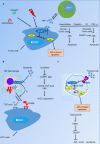
Role of DUSP1/MKP1 in tumorigenesis, tumor progression and therapy
- PMID: 27227569
- PMCID: PMC4884638
- DOI: 10.1002/cam4.772
Role of DUSP1/MKP1 in tumorigenesis, tumor progression and therapy
Abstract
Dual-specificity phosphatase-1 (DUSP1/MKP1), as a member of the threonine-tyrosine dual-specificity phosphatase family, was first found in cultured murine cells. The molecular mechanisms of DUSP1-mediated extracellular signal-regulated protein kinases (ERKs) dephosphorylation have been subsequently identified by studies using gene knockout mice and gene silencing technology. As a protein phosphatase, DUSP1 also downregulates p38 MAPKs and JNKs signaling through directly dephosphorylating threonine and tyrosine. It has been detected that DUSP1 is involved in various functions, including proliferation, differentiation, and apoptosis in normal cells. In various human cancers, abnormal expression of DUSP1 was observed which was associated with prognosis of tumor patients. Further studies have revealed its role in tumorigenesis and tumor progression. Besides, DUSP1 has been found to play a role in tumor chemotherapy, immunotherapy, and biotherapy. In this review, we will focus on the function and mechanism of DUSP1 in tumor cells and tumor treatment.
Keywords: Carcinogenesis; DUSP1/MKP1; JNK; tumor therapy.
© 2016 The Authors. Cancer Medicine published by John Wiley & Sons Ltd.
Figures

Similar articles
-
Adrenergic Stimulation of DUSP1 Impairs Chemotherapy Response in Ovarian Cancer.Clin Cancer Res. 2016 Apr 1;22(7):1713-24. doi: 10.1158/1078-0432.CCR-15-1275. Epub 2015 Nov 18. Clin Cancer Res. 2016. PMID: 26581245 Free PMC article.
-
DUSP1/MKP1 promotes angiogenesis, invasion and metastasis in non-small-cell lung cancer.Oncogene. 2011 Feb 10;30(6):668-78. doi: 10.1038/onc.2010.449. Epub 2010 Oct 4. Oncogene. 2011. PMID: 20890299
-
MAPK pathways regulation by DUSP1 in the development of osteosarcoma: Potential markers and therapeutic targets.Mol Carcinog. 2017 Jun;56(6):1630-1641. doi: 10.1002/mc.22619. Epub 2017 Feb 15. Mol Carcinog. 2017. PMID: 28112450
-
Role of Dual-Specificity Phosphatase 1 in Glucocorticoid-Driven Anti-inflammatory Responses.Front Immunol. 2019 Jun 26;10:1446. doi: 10.3389/fimmu.2019.01446. eCollection 2019. Front Immunol. 2019. PMID: 31316508 Free PMC article. Review.
-
Role of SOCS1 in tumor progression and therapeutic application.Int J Cancer. 2012 May 1;130(9):1971-80. doi: 10.1002/ijc.27318. Epub 2012 Jan 11. Int J Cancer. 2012. PMID: 22025331 Review.
Cited by
-
Oncogenic Tyrosine Phosphatases: Novel Therapeutic Targets for Melanoma Treatment.Cancers (Basel). 2020 Sep 29;12(10):2799. doi: 10.3390/cancers12102799. Cancers (Basel). 2020. PMID: 33003469 Free PMC article. Review.
-
Annurca apple polyphenol extract selectively kills MDA-MB-231 cells through ROS generation, sustained JNK activation and cell growth and survival inhibition.Sci Rep. 2019 Sep 10;9(1):13045. doi: 10.1038/s41598-019-49631-x. Sci Rep. 2019. PMID: 31506575 Free PMC article.
-
N6-methyladenosine is required for the hypoxic stabilization of specific mRNAs.RNA. 2017 Sep;23(9):1444-1455. doi: 10.1261/rna.061044.117. Epub 2017 Jun 13. RNA. 2017. PMID: 28611253 Free PMC article.
-
MiR-337-3p confers protective effect on facet joint osteoarthritis by targeting SKP2 to inhibit DUSP1 ubiquitination and inactivate MAPK pathway.Cell Biol Toxicol. 2023 Jun;39(3):1099-1118. doi: 10.1007/s10565-021-09665-2. Epub 2021 Oct 25. Cell Biol Toxicol. 2023. PMID: 34697729
-
DUSP1 interacts with and dephosphorylates VCP to improve mitochondrial quality control against endotoxemia-induced myocardial dysfunction.Cell Mol Life Sci. 2023 Jul 18;80(8):213. doi: 10.1007/s00018-023-04863-z. Cell Mol Life Sci. 2023. PMID: 37464072 Free PMC article.
References
-
- Farooq, A. , and Zhou M. M.. 2004. Structure and regulation of MAPK phosphatases. Cell. Signal. 16:769–779. - PubMed
-
- Kwak, S. P. , Hakes D. J., Martell K. J., and J. E. Dixon . 1994. Isolation and characterization of a human dual specificity protein‐tyrosine phosphatase gene. J. Biol. Chem. 269:3596–3604. - PubMed
-
- Tanoue, T. , Adachi M., Moriguchi T., and Nishida E.. 2000. A conserved docking motif in MAP kinases common to substrates, activators and regulators. Nat. Cell Biol. 2:110–116. - PubMed
Publication types
MeSH terms
Substances
LinkOut - more resources
Full Text Sources
Other Literature Sources
Research Materials
Miscellaneous

