. 2016 May 26;12(5):e1005623.
doi: 10.1371/journal.ppat.1005623. eCollection 2016 May.

A Neuronal Culture System to Detect Prion Synaptotoxicity

Affiliations

A Neuronal Culture System to Detect Prion Synaptotoxicity

Cheng Fang et al. PLoS Pathog. .

Abstract

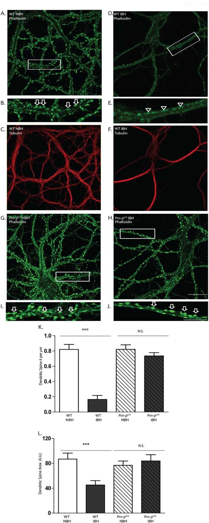
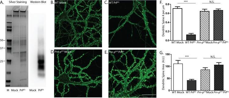
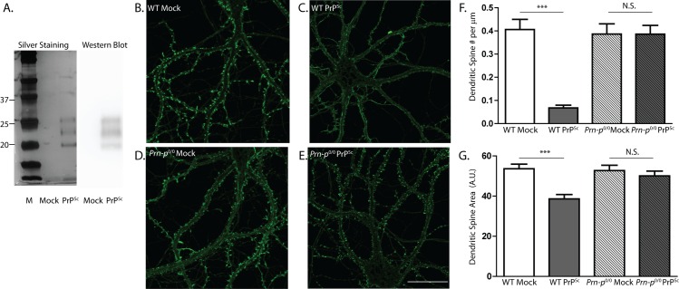
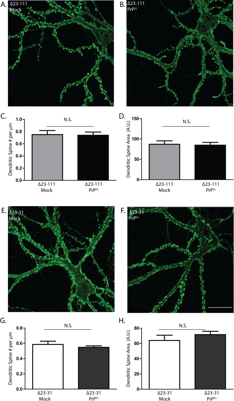
Synaptic pathology is an early feature of prion as well as other neurodegenerative diseases. Although the self-templating process by which prions propagate is well established, the mechanisms by which prions cause synaptotoxicity are poorly understood, due largely to the absence of experimentally tractable cell culture models. Here, we report that exposure of cultured hippocampal neurons to PrPSc, the infectious isoform of the prion protein, results in rapid retraction of dendritic spines. This effect is entirely dependent on expression of the cellular prion protein, PrPC, by target neurons, and on the presence of a nine-amino acid, polybasic region at the N-terminus of the PrPC molecule. Both protease-resistant and protease-sensitive forms of PrPSc cause dendritic loss. This system provides new insights into the mechanisms responsible for prion neurotoxicity, and it provides a platform for characterizing different pathogenic forms of PrPSc and testing potential therapeutic agents.


Conflict of interest statement

The authors have declared that no competing interests exist.

Figures

Fig 1
Fig 1. Scrapie-infected brain homogenate causes PrPC-dependent loss of dendritic spines.
Primary hippocampal neurons from wild-type (WT) mice (A-F) or PrP knockout (Prn-p 0/0) mice (G-J) were treated for 24 hr with brain homogenate (0.16% [w/v] final concentration) prepared from either normal mice (NBH) (A-C, G, I) or from terminally ill, scrapie-infected mice (IBH) (D-F, H, J). Neurons were then fixed and stained with Alexa 488-phalloidin (green) (A, B, D, E, G-J) to visualize F-actin, which is enriched in dendritic spines; and with anti-tubulin (red) (C, F) to visualize overall dendritic morphology. The boxed regions in panels A, D, G, and H are shown at higher magnification in panels B, E, I, and J, respectively. Arrows in panels B, I, and J point to dendritic spines, and arrowheads in panel E indicate the positions of spines that have retracted. Scale bar in panel H = 20 μm (applicable to panels A, C, D, F, G and H); scale bar in panel J = 10 μm (applicable to panels B, E, and I). Pooled measurements of spine number (K) and area (L) were collected from 16 neurons from 4 independent experiments for each genotype and each treatment. Spine number is expressed per μm length of dendrite, and spine area as the average area of an individual spine in arbitrary units (A.U.). ***p<0.001 by Student’s t-test; N.S., not significantly different. The decrease in spine area (L) reflects the fact that spines gradually shrink prior to completely disappearing; the magnitude of this effect is typically less than the reduction in the number of spines (K).
Fig 2
Fig 2. Purified PrPSc, prepared without proteases, causes PrPC-dependent spine loss.
(A) Silver stain and Western blot analysis (using anti-PrP antibody D18) of PrPSc purified from scrapie-infected brains without proteases, and mock-purified material from uninfected brains. Lane M, molecular size markers in kDa. Hippocampal neurons from wild-type (WT) mice (B, C) and PrP knockout (Prn-p 0/0) mice (D, E) were treated for 24 hr with 4.4 μg/ml of purified PrPSc (C, E), or with an equivalent amount of material mock-purified from uninfected brains (B, D). Neurons were then fixed and stained with Alexa 488-phalloidin. Scale bar in panel E = 20 μm (applicable to panels B-D). Pooled measurements of spine number (F) and area (G) were collected from 22–25 cells from 4 independent experiments. ***p<0.001 by Student’s t-test; N.S., not significantly different.
Fig 3
Fig 3. Purified PrPSc, prepared using pronase E, causes PrPC-dependent spine loss.
(A) Silver stain and Western blot analysis (using anti-PrP antibody IPC1) of PrPSc purified from scrapie-infected brains using pronase E, and mock-purified material from uninfected brains. Lane M, molecular size markers in kDa. Hippocampal neurons from wild-type (WT) mice (B, C) and PrP knockout (Prn-p 0/0) mice (D, E) were treated for 24 hr with 4.4 μg/ml of purified PrPSc (C, E), or with an equivalent amount of material mock-purified from uninfected brains (B, D). Neurons were then fixed and stained with Alexa 488-phalloidin. Scale bar in panel E = 20 μm (applicable to panels B-D). Pooled measurements of spine number (F) and area (G) were collected from 16–18 cells from 3 independent experiments. ***p<0.001 or *p<0.05 by Student’s t-test; N.S., not significantly different.
Fig 4
Fig 4. PK-digested PrPSc causes dendritic spine loss.
(A) Silver stain and Western blot (using anti-PrP antibody D18) of a PrPSc sample and a mock-purified control sample, after digestion with PK. Lane M, molecular size markers in kDa. Hippocampal neurons from wild-type (WT) mice (B, C) and PrP knockout (Prn-p 0/0) mice (D, E) were treated for 24 hr with 4.4 μg/ml of purified, PK-treated PrPSc (C, E), or with an equivalent amount of mock-purified sample (B, D). Neurons were then fixed and stained with Alexa 488-phalloidin. Scale bar in panel E = 20 μm (applicable to panels B-D). Pooled measurements of spine number (F) and area (G) were collected from 20–24 cells from 3 independent experiments. ***p<0.001 by Student’s t-test; N.S., not significantly different.
Fig 5
Fig 5. The N-terminal domain of PrPC is essential for PrPSc-induced dendritic spine loss.
Hippocampal neurons from Tg(Δ23–111) mice (A-D) and Tg(Δ23–31) mice (E-H) (both on the Prn-p 0/0 background) were treated for 24 hr with 4.4 μg/ml of PrPSc purified without proteases (B, F), or with an equivalent amount of mock-purified material from uninfected brains (A, E). Neurons were then fixed and stained with Alexa 488-phalloidin. Scale bar in panel F = 20 μm (applicable to panels A, B, E). Pooled measurements of spine number (C, G) and area (D, H) were collected from 20–24 cells from 4 independent experiments. N.S., not significantly different by Student’s t-test.

References

    1. Prusiner SB (1998) Prions. Proc Natl Acad Sci USA 95: 13363–13383. - PMC - PubMed
    1. Aguzzi A, Polymenidou M (2004) Mammalian prion biology: one century of evolving concepts. Cell 116: 313–327. - PubMed
    1. Colby DW, Prusiner SB (2011) Prions. Cold Spring Harb Perspect Biol 3: a006833 10.1101/cshperspect.a006833 - DOI - PMC - PubMed
    1. Liberski PP, Ironside JW (2004) An outline of the neuropathology of transmissible spongiform encephalopathies (prion diseases). Folia Neuropathol 42 Suppl B: 39–58. - PubMed
    1. Mallucci GR (2009) Prion neurodegeneration: starts and stops at the synapse. Prion 3: 195–201. - PMC - PubMed

Publication types

LinkOut - more resources