Comparative Study
. 2016 Sep;101(9):3459-68.
doi: 10.1210/jc.2015-4275. Epub 2016 May 26.

Enhanced Inflammatory Transcriptome in the Granulosa Cells of Women With Polycystic Ovarian Syndrome

Affiliations
Comparative Study

Enhanced Inflammatory Transcriptome in the Granulosa Cells of Women With Polycystic Ovarian Syndrome

Jaye Adams et al. J Clin Endocrinol Metab. 2016 Sep.

Abstract

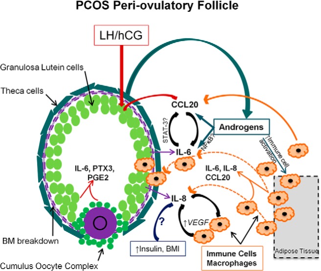
Context: Polycystic ovarian syndrome (PCOS), the most common endocrine disorder of reproductive-aged women, is associated with systemic low-grade inflammation.

Objective: We propose that increased or altered intrafollicular inflammatory reactions also occur in periovulatory follicles of PCOS patients.

Design: Gene profiling and quantitative PCR (qPCR) analyses in granulosa-lutein cells (GCs) collected from PCOS and non-PCOS women undergoing in vitro fertilization were compared with serum and follicular fluid (FF) levels of cytokines and chemokines.

Setting: This was a university-based study.

Patients: Twenty-one PCOS and 45 control patients were recruited: demographic, hormone, body mass index, and pregnancy outcomes were abstracted from patient data files.

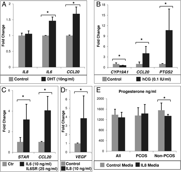
Interventions: GC cytokine/chemokine mRNAs were identified and analyzed by gene-chip microarrays/qPCR before and after culture with human chorionic gonadotropin, DHT, IL-6, or IL-8; serum/FF cytokine levels were also analyzed.

Main outcome measures: Relative serum/FF cytokine levels and GC cytokine expression before and after culture were compared and related to body mass index.

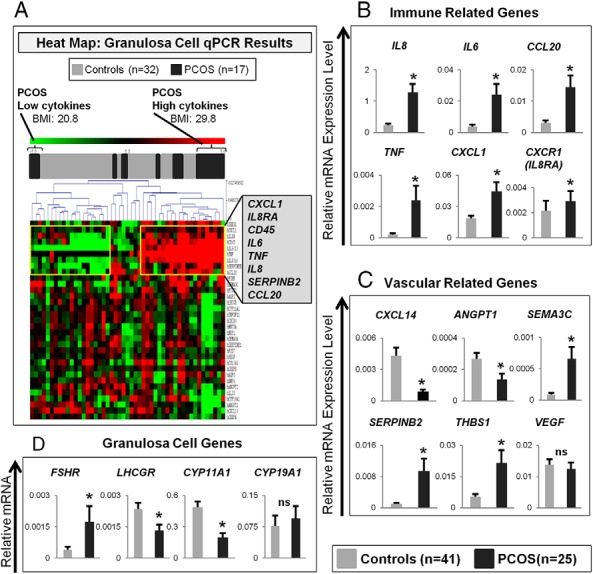
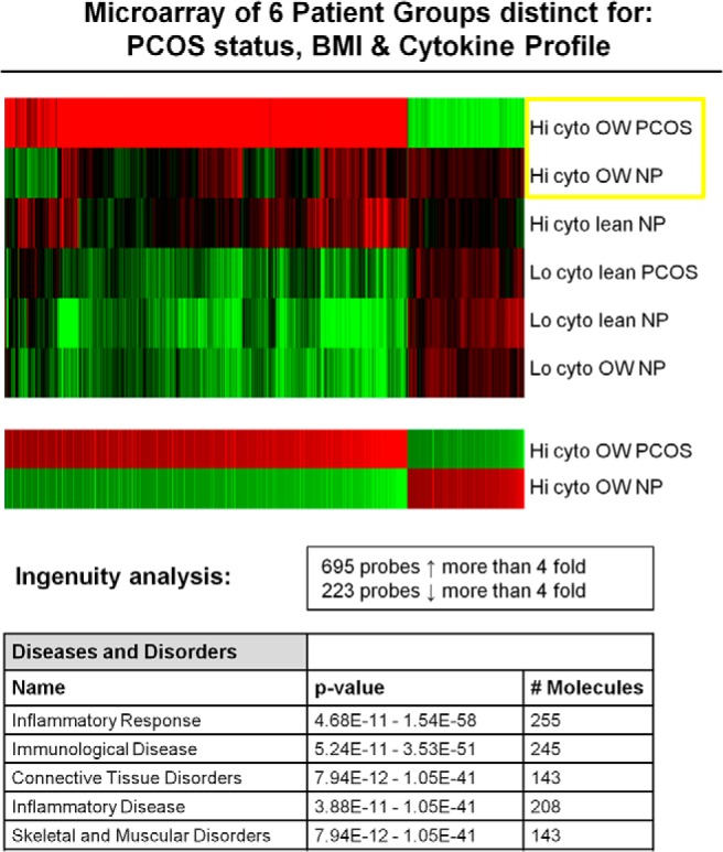
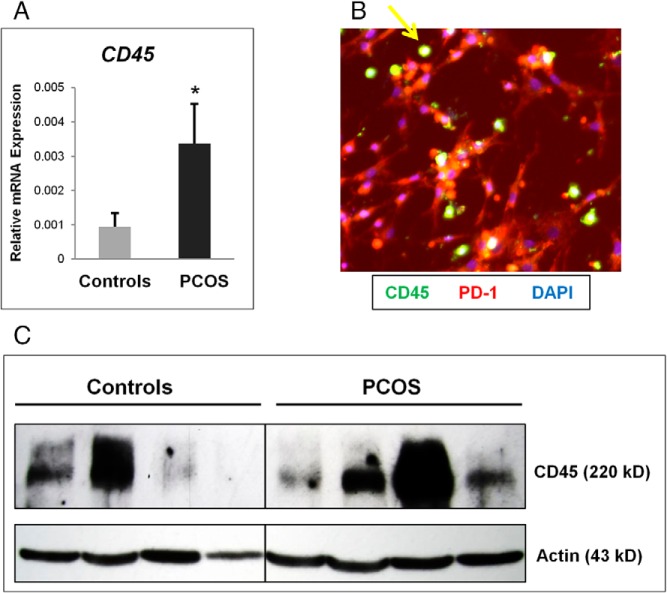
Results: The following results were found: 1) PCOS GCs express elevated transcripts encoding cytokines, chemokines, and immune cell markers, 2) based on gene profiling and qPCR analyses, obese PCOS patients define a distinct PCOS disease subtype with the most dramatic increases in proinflammatory and immune-related factors, and 3) human chorionic gonadotropin and DHT increased cytokine production in cultured GCs, whereas cytokines augmented cytokine and vascular genes, indicating that hyperandrogenism/elevated LH and obesity in PCOS women augment intrafollicular cytokine production.

Conclusions: Intrafollicular androgens and cytokines likely comprise a local regulatory loop that impacts GC expression of cytokines and chemokines and the presence of immune cells; this loop is further enhanced in the obese PCOS subtype.


Figures

Figure 1.
Figure 1.
A, Heat map of relative qPCR results. B–D, Relative mRNA expression of genes in luteinized granulosa cell collections of control compared with PCOS women. B, Immune-related genes. C, Vascular-related genes. D, Genes specific to granulosa cells. Statistical comparisons were made with the Mann-Whitney test. *, P < .05. ns, not significant.
Figure 2.
Figure 2.
This microarray figure depicts the six groups organized from highest to lowest discordance. The two overweight high-cytokine groups (PCOS vs non-PCOS) are compared separately together in the lower figure, which shows a greater than 4-fold alteration in the expression for 918 gene probes between those two groups. The top five categories of increased gene regulation via the Ingenuity analysis are shown in the lower table. BMI, body mass index; cyto, cytokine; Hi, high; Lo, low; NP, samples from normoovulatory patients; PCOS, samples from patients with PCOS.
Figure 3.
Figure 3.
CD45 expression is increased in PCOS compared with control granulosa cell collections. A, CD45 mRNA expression is increased in granulosa cell collections from the PCOS compared with the control group. *, P < .05. B, Immunofluorescent identification of leukocytes (CD45) in PCOS granulosa cells (PD-1) cultured on coverslips for 5 days. Yellow arrow indicates CD45+ leukocyte cell. C, Western blot of granulosa cell lysate in representative control and PCOS samples, normalized to β-actin levels. Note the marked variation of CD45 protein levels and mRNA expression common in individual patient samples but overall increased in the PCOS group as a whole.
Figure 4.
Figure 4.
Relative mRNA expression in granulosa cells cultured in vitro with the indicated treatment for 24 hours in serum-free media. For A–D, data are shown as fold change (mean ± SEM) relative to control (vehicle only) group, which is equivalent to 1. For A–E, data were analyzed by a Wilcoxon's signed rank test. *, P < .05. n, number of separate culture experiments with different patient samples. A, DHT treatment (n = 12). B, hCG treatment for 24 hours in serum-free media after 5 days of culture (n = 5). C, Treatment with IL-6 and IL-6 soluble receptor (IL6SR) (n = 9). D, IL-8 treatment (n = 9). E, Progesterone production as measured in spent media from IL-8-treated granulosa cells in culture (n = 10). Progesterone levels in control were compared with IL-8-treated wells and were further stratified by clinical grouping. Whereas cells from PCOS women (n = 4) demonstrated a small, nonsignificant increase in progesterone production, those from control women (n = 6) showed a significant decrease in progesterone production.
Figure 5.
Figure 5.
Working model of enhanced cytokine production in the PCOS periovulatory follicle. Cytokines and chemokines are secreted by GCs and are physiological components of the ovulatory response. Elevated local androgens in the PCOS follicle may increase IL6 and CCL20 expression, which can then enhance immune cell activation and recruitment to the area. These recruited immune cells then further contribute to the production of local inflammatory molecules, possibly affecting both angiogenic and steroidogenic pathways. IL8 may be increased by systemic factors associated with elevated body mass index and insulin levels and may also participate in a synergistic-positive feedback loop with immune cells in the production of VEGF and other proangiogenic factors. BM, basement membrane.

References

    1. Norman RJ, Brannstrom M. Cytokines in the ovary: pathophysiology and potential for pharmacological intervention. Pharmacol Ther. 1996;69(3):219–236. - PubMed
    1. Hellberg P, Thomsen P, Janson PO, Brannstrom M. Leukocyte supplementation increases the luteinizing hormone-induced ovulation rate in the in vitro-perfused rat ovary. Biol Reprod. 1991;44(5):791–797. - PubMed
    1. Brannstrom M, Bonello N, Wang LJ, Norman RJ. Effects of tumour necrosis factor α (TNF α) on ovulation in the rat ovary. Reprod Fertil Dev. 1995;7(1):67–73. - PubMed
    1. Brannstrom M, Bonello N, Norman RJ, Robertson SA. Reduction of ovulation rate in the rat by administration of a neutrophil-depleting monoclonal antibody. J Reprod Immunol. 1995;29(3):265–270. - PubMed
    1. Smolikova K, Mlynarcikova A, Scsukova S. Role of interleukins in the regulation of ovarian functions. Endocr Regul. 2012;46(4):237–253. - PubMed

Publication types

MeSH terms