. 2016 Jun;26(3):156-65.
doi: 10.1089/nat.2015.0599. Epub 2016 May 26.

Improved Synthesis and In Vitro Evaluation of an Aptamer Ribosomal Toxin Conjugate

Affiliations

Improved Synthesis and In Vitro Evaluation of an Aptamer Ribosomal Toxin Conjugate

Linsley Kelly et al. Nucleic Acid Ther. 2016 Jun.

Abstract

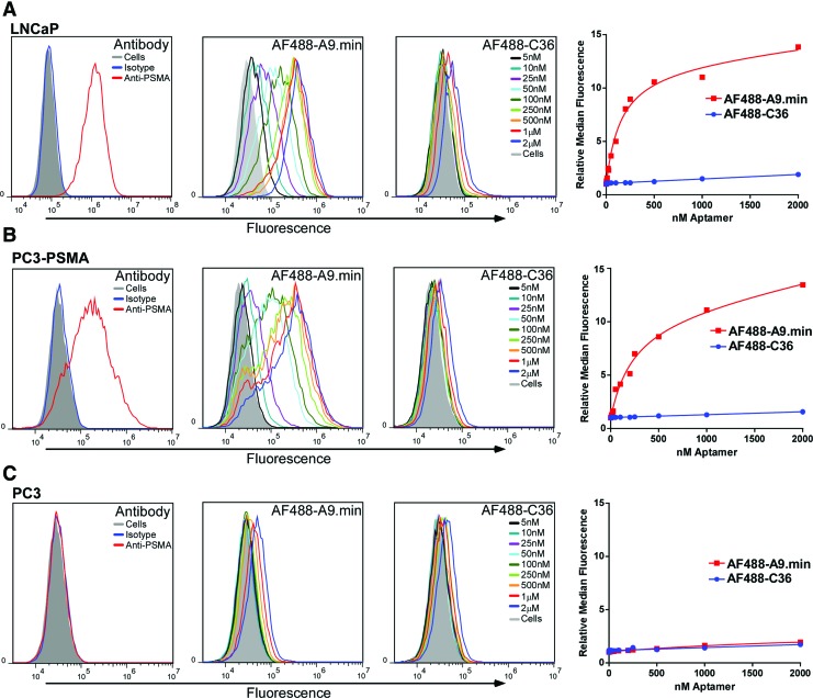
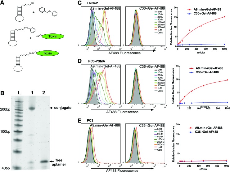
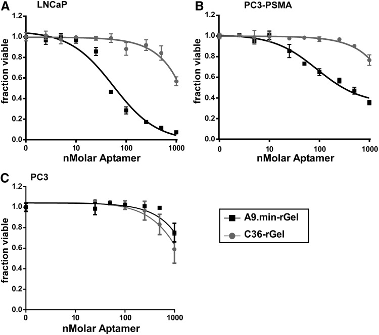
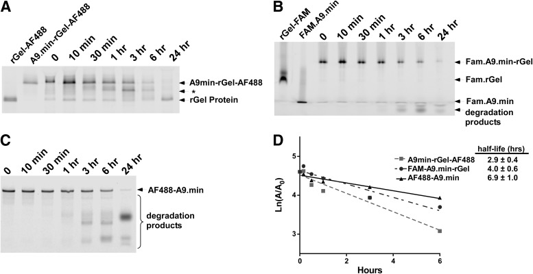
Delivery of toxins, such as the ricin A chain, Pseudomonas exotoxin, and gelonin, using antibodies has had some success in inducing specific toxicity in cancer treatments. However, these antibody-toxin conjugates, called immunotoxins, can be bulky, difficult to express, and may induce an immune response upon in vivo administration. We previously reported delivery of a recombinant variant of gelonin (rGel) by the full-length prostate-specific membrane antigen (PSMA) binding aptamer, A9, to potentially circumvent some of these problems. Here, we report a streamlined approach to generating aptamer-rGel conjugates utilizing a chemically synthesized minimized form of the A9 aptamer. Unlike the full-length A9 aptamer, this minimized variant can be chemically synthesized with a 5' terminal thiol. This facilitates the large scale synthesis and generation of aptamer toxin conjugates linked by a reducible disulfide linkage. Using this approach, we generated aptamer-toxin conjugates and evaluated their binding specificity and toxicity. On PSMA(+) LNCaP prostate cancer cells, the A9.min-rGel conjugate demonstrated an IC50 of ∼60 nM. Additionally, we performed a stability analysis of this conjugate in mouse serum where the conjugate displayed a t1/2 of ∼4 h, paving the way for future in vivo experiments.


Figures

<b>FIG. 1.</b>
FIG. 1.
Cell binding and PSMA specificity analysis of the anti-PSMA aptamer A9.min. (A) LNCaP, (B) PC3-PSMA, and (C) PC3 cells were incubated with an anti-PSMA antibody, and isotype controls or increasing concentrations of AF488-labeled A9.min or a nonfunctional aptamer control, AF488-labeled C36. LNCaP and PC3-PSMA cells both express PSMA. PC3 cells are a PSMA-negative cell line as confirmed by antibody staining. Cell staining experiments were performed in growth media containing 10% FBS supplemented with 1 mg/mL ssDNA as a non-specific blocking agent. Incubations were performed for 1 h after which the cells were washed to remove unbound aptamers, trypsinized from the plate and analyzed by flow cytometry. The identity and concentration of each plot in the histograms are as indicated. For each data set, we also generated a plot of the relative median fluorescence intensity versus concentration, which was used to determine the apparent binding constant (appKd) on each cell type. The appKd values are reported in Table 1. FBS, fetal bovine serum; PSMA, prostate-specific membrane antigen. Color images available online at www.liebertpub.com/nat
<b>FIG. 2.</b>
FIG. 2.
Synthesis and binding characteristics of A9.min-rGel. (A) Synthesis scheme for the generation of aptamer-toxin conjugates using a 5′ thiol modified aptamer. (i) The reduced aptamer bearing a 5′ thiol is activated by reaction with dithiodipyridine (DTDP). (ii) The activated, thiol reactive aptamer is incubated with target protein bearing a single Cys residue. (iii) The resulting conjugate bears a reducible disulfide linkage. (B) Electrophoretic analysis of purified A9.min-rGel before (lane 1) and after treatment with the reducing agent TCEP (lane 2). (C) LNCaP, (D) PC3-PSMA, and (E) PC3 cells were incubated with increasing concentrations of an A9.min-rGel conjugate in which the protein was labeled with AF488 (A9.min-rGel-AF488) or a nonfunctional aptamer conjugate control (C36-rgel-AF488). LNCaP and PC3-PSMA cells both express PSMA. PC3 cells are a PSMA-negative cell line. Cell staining experiments were performed in growth media containing 10% FBS supplemented with 1 mg/mL ssDNA as a nonspecific blocking agent. Incubations were performed for 1 h after which the cells were washed to remove unbound aptamers, trypsinized from the plate, and analyzed by flow cytometry. The identity and concentration of each plot in the histograms are as indicated. For each data set, we also generated a plot of the relative median fluorescence intensity versus concentration which was used to determine the apparent binding constant (appKd) on each cell type. The appKd values are reported in Table 1. Color images available online at www.liebertpub.com/nat
<b>FIG. 3.</b>
FIG. 3.
Cell killing properties of A9.min-rGel. (A) LNCaP, (B) PC3-PSMA, and (C) PC3 cells were incubated with increasing concentrations of A9.min-rGel or a nonfunctional aptamer conjugate control C36-rGel. LNCaP and PC3-PSMA cells both express PSMA. PC3 cells are a PSMA-negative cell line. Assays were performed in growth media containing 10% FBS supplemented with 1 mg/mL ssDNA as a nonspecific blocking agent. Conjugates were added to cells at increasing concentrations and incubated for 5 days after which the fraction of viable cells remaining was determined. The IC50 values for each curve are reported in Table 1.
<b>FIG. 4.</b>
FIG. 4.
Serum stability of A9.min and A9.min-rGel conjugates. (A) Analysis of A9.min-rGel conjugate in which the protein was labeled with AF488 (A9.min-rGel-AF488). (B) Analysis of an A9.min-rGel conjugate in which the aptamer was labeled at the 3′ end with FAM (FAM.A9.min-rGel). (C) Analysis of A9.min bearing a 5′ AF488 and a 3′ inverted dT residue (AF499-A9.min). Assays were performed in 100% fresh mouse serum for the time indicated. The identity of each band is as indicated. rGel-AF488 and rGel-FAM are fluorescently labeled rGel standards. FAM.A9.min is the 3′ FAM labeled A9.min aptamer and * indicates the position of the degradation product that displays intermediate electrophoretic mobility between that of the full-length conjugate and the protein alone. Sample workup before electrophoresis was performed as described in the Methods and Materials section. (D) Serum stability was determined from a plot of the area corresponding to the full-length conjugate or aptamer on the gel. The half-life for each sample is as indicated.

Similar articles

Cited by

References

    1. Pastan I, Hassan R, FitzGerald DJ. and Kreitman RJ. (2007). Immunotoxin treatment of cancer. Annu Rev Med 58:221–237 - PubMed
    1. Ray P, Cheek MA, Sharaf ML, Li N, Ellington AD, Sullenger BA, Shaw BR. and White RR. (2012). Aptamer-mediated delivery of chemotherapy to pancreatic cancer cells. Nucleic Acid Ther 22:295–305 - PMC - PubMed
    1. Zhao N, Pei SN, Qi J, Zeng Z, Iyer SP, Lin P, Tung CH. and Zu Y. (2015). Oligonucleotide aptamer-drug conjugates for targeted therapy of acute myeloid leukemia. Biomaterials 67:42–51 - PMC - PubMed
    1. Huang YF, Shangguan D, Liu H, Phillips JA, Zhang X, Chen Y. and Tan W. (2009). Molecular assembly of an aptamer-drug conjugate for targeted drug delivery to tumor cells. Chembiochem 10:862–868 - PMC - PubMed
    1. Farokhzad OC, Jon S, Khademhosseini A, Tran TN, Lavan DA. and Langer R. (2004). Nanoparticle-aptamer bioconjugates: a new approach for targeting prostate cancer cells. Cancer Res 64:7668–7672 - PubMed

MeSH terms

LinkOut - more resources