The effect of amifostine on differentiation of the human megakaryoblastic Dami cell line
- PMID: 27228575
- PMCID: PMC4884634
- DOI: 10.1002/cam4.759
The effect of amifostine on differentiation of the human megakaryoblastic Dami cell line
Abstract
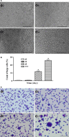
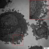
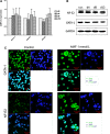
Amifostine is a cytoprotective drug that was initially used to control and treat nuclear radiation injury and is currently used to provide organ protection in cancer patients receiving chemotherapy. Clinical studies have also found that amifostine has some efficacy in the treatment of cytopenia caused by conditions such as myelodysplastic syndrome and immune thrombocytopenia, both of which involve megakaryocyte maturation defects. We hypothesized that amifostine induced the differentiation of megakaryocytes and investigated this by exposing the human Dami megakaryocyte leukemia cell line to amifostine (1 mmol/L). After 12 days of amifostine exposure, optical microscopy showed that the proportion of Dami cells with diameters >20 μm had increased to 24.63%. Transmission electron microscopy identified the development of a platelet demarcation membrane system, while flow cytometry detected increased CD41a expression and decreased CD33 expression on the Dami cell surface. Ploidy analysis found that the number of polyploid cells with >4N DNA content increased to 27.96%. We did not detect any elevation in the mRNA or protein levels of megakaryocytic differentiation-associated transcription factors GATA-binding factor 1 (GATA-1) and nuclear factor, erythroid 2 (NF-E2), but nuclear import assay revealed an increased nuclear translocation of these proteins. These findings indicate that amifostine induced the differentiation of Dami cells into mature megakaryocytes via a mechanism involving increased nuclear translocation of the transcription factors, NF-E2 and GATA-1.
Keywords: Amifostine; CD41a; DNA ploidy; Dami cells; transcription factor.
© 2016 The Authors. Cancer Medicine published by John Wiley & Sons Ltd.
Figures


References
-
- Valeriote, F. , and Tolen S.. 1982. Protection and potentiation of nitrogen mustard cytotoxicity by WR‐2721. Cancer Res. 42:4330–4331. - PubMed
-
- Capizzi, R. L. , Scheffler B. J., and Schein P. S.. 1993. Amifostine‐mediated protection of normal bone marrow from cytotoxic chemotherapy. Cancer 72(11 Suppl.):3495–3501. - PubMed
-
- Kanter, M. , Topcu‐Tarladacalisir Y., and Uzal C.. 2011. Role of amifostine on acute and late radiation nephrotoxicity: a histopathological study. In Vivo. 25:77–85. - PubMed
-
- List, A. F. , Heaton R., Glinsmann‐Gibson B., and Capizzi R. L.. 1998. Amifostine stimulates formation of multipotent and erythroid bone marrow progenitors. Leukemia 12:1596–1602. - PubMed
Publication types
MeSH terms
Substances
LinkOut - more resources
Full Text Sources
Other Literature Sources

