. 2016 Jun 14;7(24):35577-35591.
doi: 10.18632/oncotarget.9590.

Impaired macrophage autophagy induces systemic insulin resistance in obesity

Affiliations

Impaired macrophage autophagy induces systemic insulin resistance in obesity

Young-Ho Kang et al. Oncotarget. .

Abstract

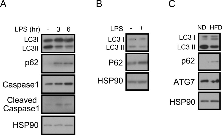
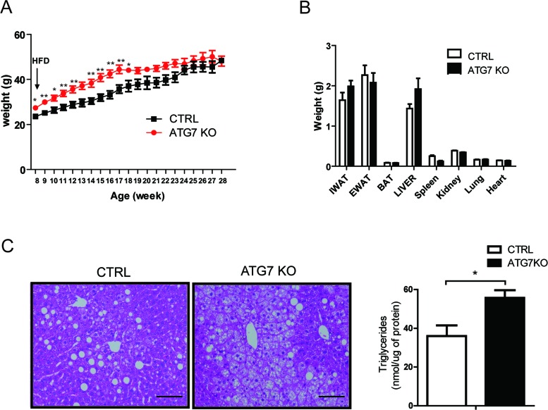
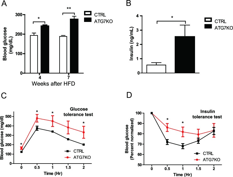
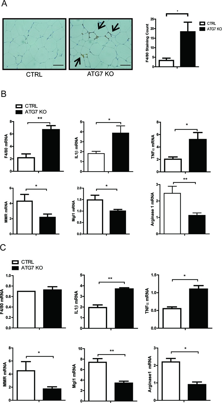
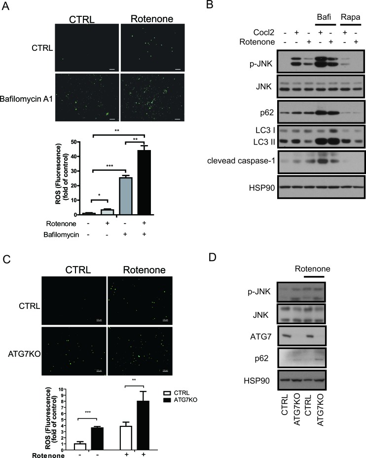
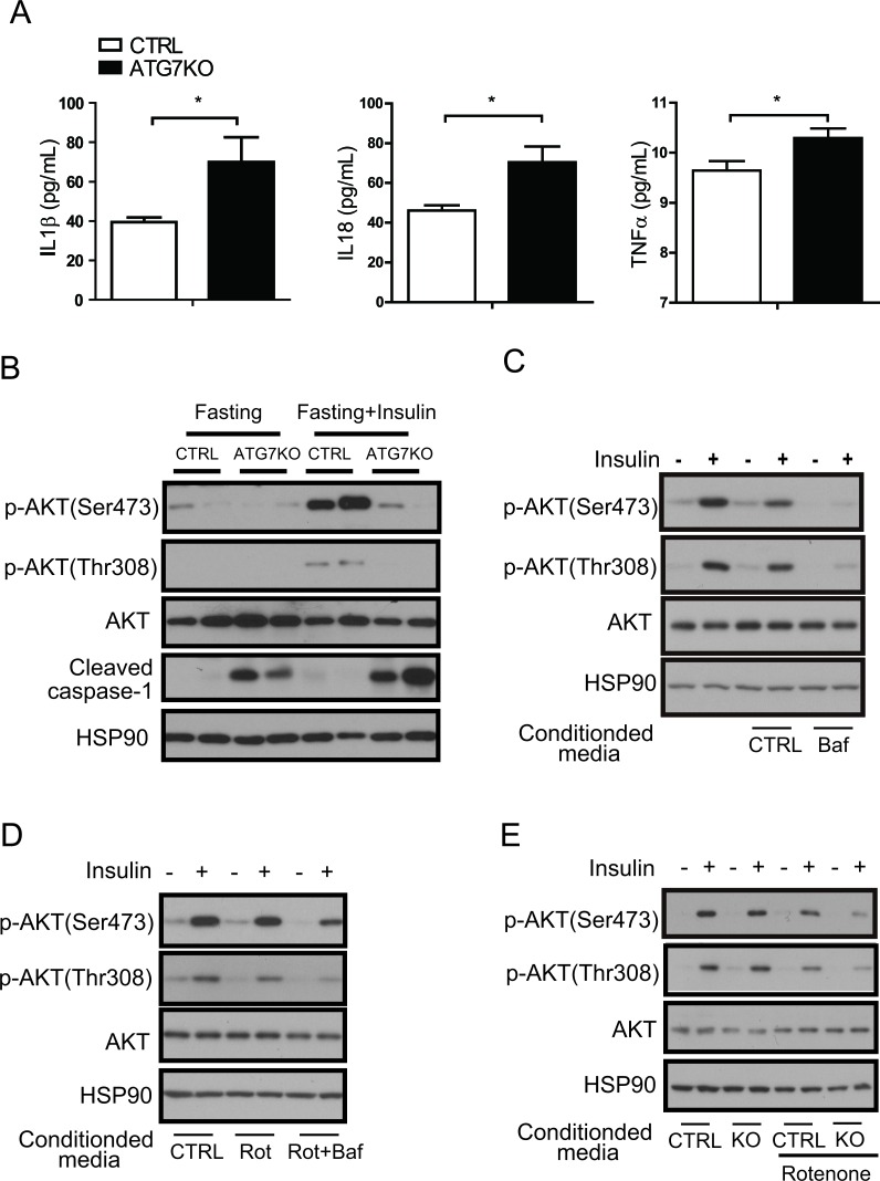
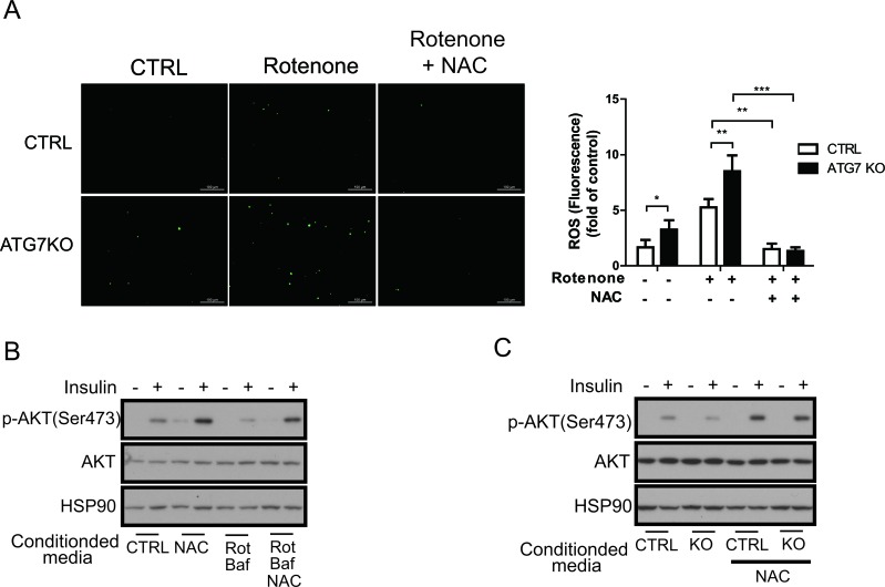
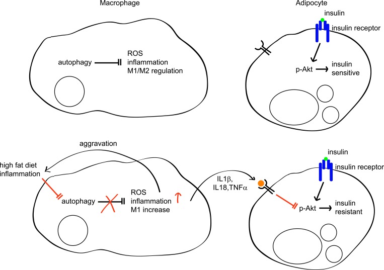
Obesity-induced insulin resistance and diabetes are significantly associated with infiltrates of inflammatory cells in adipose tissue. Previous studies recognized the involvement of autophagy in the regulation of metabolism in multiple tissues, including β-cells, hepatocytes, myocytes, and adipocytes. However, despite the importance of macrophages in obesity-induced insulin resistance, the role of macrophage autophagy in regulating insulin sensitivity is seldom addressed. In the present study, we show that macrophage autophagy is important for the regulation of systemic insulin sensitivity. We found that macrophage autophagy is downregulated by both acute and chronic inflammatory stimuli, and blockade of autophagy significantly increased accumulation of reactive oxygen species (ROS) in macrophages. Macrophage-specific Atg7 knockout mice displayed a shift in the proportion to pro-inflammatory M1 macrophages and impairment of insulin sensitivity and glucose homeostasis under high-fat diet conditions. Furthermore, inhibition of ROS in macrophages with antioxidant recovered adipocyte insulin sensitivity. Our results provide evidence of the underlying mechanism of how macrophage autophagy regulates inflammation and insulin sensitivity. We anticipate our findings will serve as a basis for development of therapeutics for inflammatory diseases, including diabetes.

Keywords: Pathology Section; adipose tissue macrophage; autophagy; insulin resistance; obesity; reactive oxygen species.


Conflict of interest statement

The authors declare that there is no conflict of interest associated with this manuscript.

Figures

Figure 1
Figure 1. Impaired autophagy and increased IL1β in LPS-treated macrophages
A. Western blots showing that LC3-II is decreased, p62-sequestosome-1 is increased, and the cleaved form of caspase-1 is increased in LPS (1 ng/mL)-treated BMDMs from C57BL6 mice. HSP90 was loaded as an internal control. B. Western blots showing that LC3-II is decreased and p62 is increased in the LPS-treated Raw264.7 macrophage cell line. C. Western blots showing that LC3-II conversion is decreased and p62 accumulation is increased in peritoneal macrophages from 60% HFD-fed obese mice compared with macrophages from normal chow diet (ND)-fed mice.
Figure 2
Figure 2. Macrophage-specific Atg7KO mice develop hepatic steatosis under HFD conditions
A. Atg7fl/fl-LysMCre+/− mice (Atg7KO) (n = 8) and LysMCre+/− mice (control) (n = 6) were fed a 60% HFD, and their growth was monitored by weighing weekly. B. Twenty weeks after administering a 60% HFD, the tissues from Atg7KO (n = 8) and control mice (n = 6) were dissected and weighed. C. Liver sections from Atg7KO and control mice were prepared and stained with hematoxylin and eosin staining. Scale bar, 100 μm (left panel). The triglyceride content of liver tissues from control and Atg7KO mice is shown in the right panel.
Figure 3
Figure 3. Impaired glucose homeostasis in macrophage-specific Atg7KO mice under diet-induced obesity conditions
A. Circulating blood glucose levels in Atg7KO and control mice during a 60% HFD challenge. Circulating glucose levels were monitored in 5-h-fasted (weeks 4 and 7 after HFD) mice. B. Circulating insulin levels of Atg7KO (n = 6) and control (n = 6) mice were measured using ELISA. C. GTT and D. ITT (right) results for Atg7KO (n = 8) and control (n = 6) mice.
Figure 4
Figure 4. Increased proinflammatory macrophages in the adipose tissue of macrophage-specific Atg7KO mice
A. Immunohistochemistry showing the increased infiltration of F4/80-positive macrophages in the adipose tissues of macrophage-specific Atg7KO mice. B. qPCR analysis showing that the F4/80, IL1β, and TNFα mRNA levels increased and the MMR, Mgl1, and arginase1 mRNA levels decreased in the adipose tissues of Atg7KO mice. C. qPCR analysis showing that IL1β and TNFα transcripts were upregulated and that MMR, Mgl1, and arginase1 mRNAs were downregulated in the peritoneal macrophages of Atg7KO mice.
Figure 5
Figure 5. Increased ROS levels in autophagy-impaired macrophages
A. ROS staining of Raw264.7 cells with DCF-DA dye revealed that the ROS levels increased in rotenone-treated or bafilomycin A1-treated cells. B. Western blots showing that phosphorylated JNK (p-JNK) and cleaved caspase-1 levels were increased in CoCl2-treated or rotenone-treated Raw264.7 cells and further increased in bafilomycin A1-treated cells and attenuated by rapamycin treatment. C. ROS staining of peritoneal macrophages with DCF-DA dye showing that the ROS level is increased in the Atg7KO mice and further increased by rotenone treatment. D. Western blots showing that the phosphorylation of JNK is elevated in the peritoneal macrophages of Atg7KO mice.
Figure 6
Figure 6. A macrophage autophagy block disrupts insulin signaling in adipose tissues
A. Circulating IL1β, IL18, and TNFα levels in Atg7KO (n = 6-8) and control mice (n = 6-7) were examined by Elisa. B. White adipose tissue was collected after fasting (with or without insulin injection) from Atg7KO and control mice, and cleaved caspase-1 and insulin signaling were analyzed by western blot. C. Conditioned media collected from Raw264.7 cells treated with bafilomycin A1 and D. conditioned media collected from Raw264.7 cells treated with rotenone (or rotenone and bafilomycinA1) were applied to 3T3-L1 cells with or without insulin, and insulin signaling was then examined by western blot analysis. E. Conditioned media were collected from the peritoneal macrophages of the control and Atg7KO mice and applied to the 3T3-L1 cell line. The insulin signaling pathway was then examined by western blot analysis.
Figure 7
Figure 7. Macrophage ROS are required for the regulation of insulin signaling
A. Peritoneal macrophages from control and Atg7KO mice were treated with rotenone with or without NAC. ROS level measured by DCF-DA staining shows that rotenone-mediated ROS generation is inhibited in peritoneal macrophages from both control and Atg7KO mice. B. Conditioned media were collected from Raw264.7 cells treated with NAC, rotenone, or bafilomycin A1 as indicated and applied to 3T3-L1 cells with or without insulin. Insulin signaling was examined by western blot analysis. C. Conditioned media collected from peritoneal macrophages of control and Atg7KO mice treated with NAC as indicated were applied to 3T3-L1 cells with or without insulin treatment, and insulin signaling was examined by western blot analysis.
Figure 8
Figure 8. Regulation of insulin sensitivity by autophagy in macrophages
In normal conditions, autophagy regulates inflammation, ROS levels, and M1/M2 populations in macrophages. In this context, adipocytes are sensitive to insulin. However, in inflammatory conditions such as diabetes or HFD, autophagy is impaired in macrophages and its regulation of ROS, inflammation, and the M1/M2 population is also impaired, which further aggravates inflammation and autophagy impairment. Concomitantly, increased inflammatory cytokines such as IL1β, IL18, and TNFα inhibit Akt, which impairs insulin signaling, resulting in insulin resistance.

References

    1. Ginsberg HN, Huang LS. The insulin resistance syndrome: impact on lipoprotein metabolism and atherothrombosis. Journal of cardiovascular risk. 2000;7:325–331. - PubMed
    1. Weisberg SP, McCann D, Desai M, Rosenbaum M, Leibel RL, Ferrante AW., Jr Obesity is associated with macrophage accumulation in adipose tissue. The Journal of clinical investigation. 2003;112:1796–1808. - PMC - PubMed
    1. Xu H, Barnes GT, Yang Q, Tan G, Yang D, Chou CJ, Sole J, Nichols A, Ross JS, Tartaglia LA, Chen H. Chronic inflammation in fat plays a crucial role in the development of obesity-related insulin resistance. The Journal of clinical investigation. 2003;112:1821–1830. - PMC - PubMed
    1. Heilbronn LK, Campbell LV. Adipose tissue macrophages, low grade inflammation and insulin resistance in human obesity. Current pharmaceutical design. 2008;14:1225–1230. - PubMed
    1. Wellen KE, Hotamisligil GS. Inflammation, stress, and diabetes. The Journal of clinical investigation. 2005;115:1111–1119. - PMC - PubMed

MeSH terms