. 2016 May 27:7:11694.
doi: 10.1038/ncomms11694.

Pre-bilaterian origin of the blastoporal axial organizer

Affiliations

Pre-bilaterian origin of the blastoporal axial organizer

Yulia Kraus et al. Nat Commun. .

Abstract

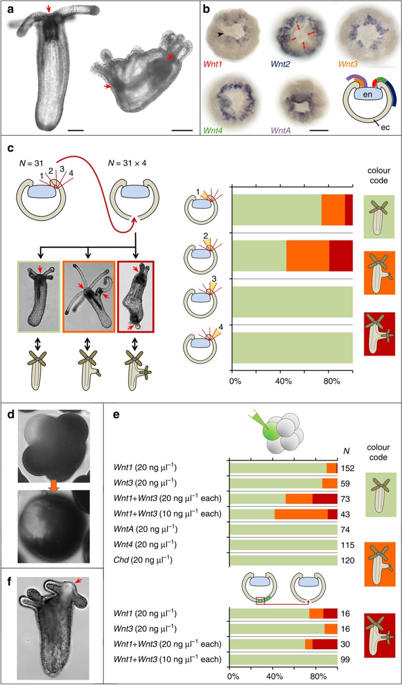
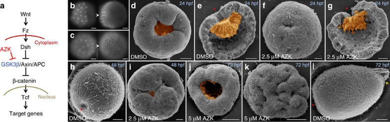
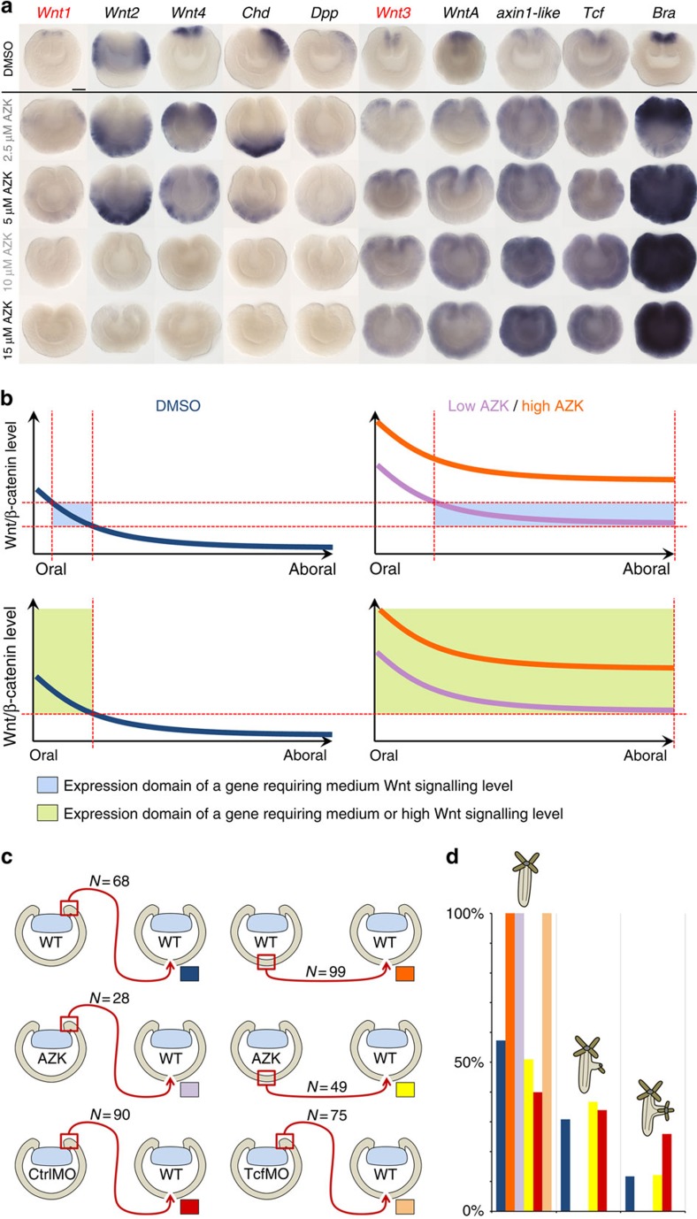
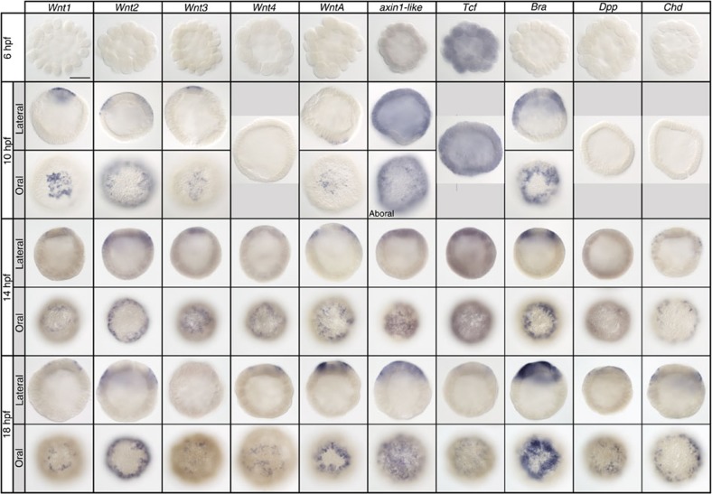
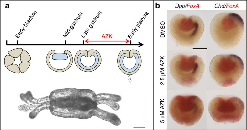
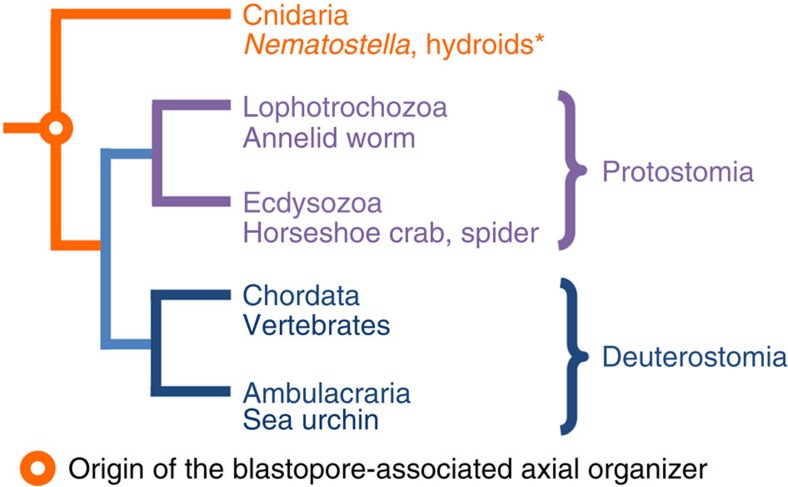
The startling capacity of the amphibian Spemann organizer to induce naïve cells to form a Siamese twin embryo with a second set of body axes is one of the hallmarks of developmental biology. However, the axis-inducing potential of the blastopore-associated tissue is commonly regarded as a chordate feature. Here we show that the blastopore lip of a non-bilaterian metazoan, the anthozoan cnidarian Nematostella vectensis, possesses the same capacity and uses the same molecular mechanism for inducing extra axes as chordates: Wnt/β-catenin signaling. We also demonstrate that the establishment of the secondary, directive axis in Nematostella by BMP signaling is sensitive to an initial Wnt signal, but once established the directive axis becomes Wnt-independent. By combining molecular analysis with experimental embryology, we provide evidence that the emergence of the Wnt/β-catenin driven blastopore-associated axial organizer predated the cnidarian-bilaterian split over 600 million years ago.


Figures

Figure 1
Figure 1. Ectopic expression of Wnt1 and Wnt3 induces ectopic body axes.
(a) CRISPR-Cas9 knockout of APC results in the formation of ectopic tentacles and pharynges in F0. Left: wild type polyp; right: mosaic mutant polyp. Red arrows—pharynges. (b) Five ectodermal Wnt genes are expressed at mid-gastrula, however, Wnt2 is expressed at a distance to the bend of the blastopore lip (red double-headed arrows). Gene names are colour-coded as on the scheme showing a lateral view on a mid-gastrula with ectodermal Wnt expression domains depicted as coloured lines. en—invaginating endoderm, ec—ectoderm. Black arrowhead points at the bend of the blastopore lip. (c) Results of transplantation of four sequential blastopore lip fragments from donor gastrulae (N=31) to four different recipient gastrulae (N=31 × 4). Possible developmental outcomes: no axis duplication (green bars on graph), incomplete axis duplication (an outgrowth with tentacles and, sometimes, pharynx but without mesenteries; orange bars on graph), complete axis duplication (two contractile axes with head structures; red bars on graph). Red arrows—pharynges. Fragment 1 and fragment 2, closest to the bend of the blastopore lip, are inductive. (d) Co-injection of a plasmid with EF1α promoter driving the expression of a gene of interest and Dextran-Alexa488 into single cells in 8–16 cell stage embryos results in formation of a coherent patch of fluorescent cells. (e) Some embryos injected into single blastomeres at 8–16 cell stage with Wnt1 or Wnt3 or with both these Wnt's develop ectopic axes. WntA, Wnt4 and Chd never induce a second axis. Fluorescent aboral ectoderm from Wnt1 and Wnt3 injected embryos also acquires inductive capacity. The colour code of the bars is the same as on (c). (f) If EF1a::mOrange2 is co-injected with the Wnt plasmids, mOrange2 fluorescence is observed in the induced secondary head (red arrow). Scale bars: 100 μm.
Figure 2
Figure 2. Early AZK treatment results in formation of severely oralized embryos.
(a) AZK upregulates Wnt/β-catenin signaling by inhibiting GSK3β. (b) Nuclear translocation of β-catenin-venus fusion protein on one side in 6 and 9 h post fertilization (hpf) embryos injected with β-catenin-venus mRNA. (c) Nuclear β-catenin-venus in all cells at 6 and 9 hpf of AZK treated blastulae. (d,e) SEM of control 24 hpf mid-gastrulae; oral (d) and lateral (e) views. Note large blastopore and pre-endodermal plate. (f,g) Small blastopore and pre-endodermal plate on SEM of mid-gastrulae after early treatment with 2.5 μM AZK. Gastrulae on (e) and (g) were split into halves to make inner structures visible. (h,i) Control 48 hpf planula (h lateral view) and a planula subjected to the early 2.5 μM AZK treatment (i, oral view). In treated planula, blastopore starts to re-open and small pits appear throughout the surface. (jl) oral (j) and aboral (k) views of the 72 hpf planula after early 5 μM AZK treatment and of the control 72 hpf planula (l). The oral surface of the treated embryo carries a re-opened blastopore (j), the aboral surface shows multiple folds and holes. The embryo fails to elongate and form aboral structures (e.g., apical tuft - yellow arrowhead on (l)). Scale bars: 30 μm. Endoderm highlighted orange. Red asterisks on lateral views denote blastopores.
Figure 3
Figure 3. Organizer capacity shifts to follow changes in Wnt1 and Wnt3 expression.
(a) Expression of Wnt1, Wnt2, Wnt4, Chd and Dpp expands aborally while clearing from the oral domain and disappears with the increasing AZK concentrations; expression of Wnt3, axin1-like, Tcf and Bra expands globally and reaches saturation. Blastopores point up; scale bar: 50 μm. (b) Model of expression of a gene activated at medium Wnt/β-catenin signaling levels (upper row; blue highlight represents the range of Wnt/β-catenin signaling levels when the gene can be expressed) versus a gene activated at medium and high Wnt/β-catenin signaling levels (lower row; green highlight represents the range of Wnt/β-catenin signaling levels when the gene can be expressed) in the AZK treatments. Blue curve: wild type level of Wnt/β-catenin signaling along the oral-aboral axis. Lilac curve: Wnt/β-catenin signaling level in a low concentration of AZK. Orange curve: Wnt/β-catenin signaling level in a high concentration of AZK. Red dashed lines mark the Wnt/β-catenin signaling thresholds and the corresponding positions on the oral-aboral axis where these thresholds are reached. (c,d) Scheme (c) and developmental outcomes (d) of transplantation of wild type (wt) and 2.5  μM AZK treated (AZK) blastopore lips and aboral ectoderm fragments as well as blastopore lips of the CtrlMO and TcfMO injected gastrulae into wild type recipient gastrulae. Coloured rectangles on (c) correspond to the colours of the bars on (d). Only untreated or CtrlMO blastopore lips and AZK treated aboral ectoderm fragments are inductive.
Figure 4
Figure 4. Establishment of the oral inductive territory in Nematostella.
At 10 hpf Wnt1, Wnt2, Wnt3 and WntA are the first Wnt genes to be expressed. Wnt2 expression starts as a ring excluding the presumptive pre-endodermal plate. Wnt1, Wnt3 and WntA expression starts as a patch in the presumptive pre-endodermal plate and by 14–18 hpf begins to form a ring around it. Brachyury expression is first detectable as a ring at 10 hpf. Wnt4, Dpp and Chd start to be expressed in a ring around the pre-endodermal plate by 14 hpf. On lateral views, animal/oral ends of the embryos point up. Scale bar: 100 μm.
Figure 5
Figure 5. Late AZK treatment affects the oral-aboral but not the directive axis.
(a) Scheme of late AZK treatment and a resulting double-headed primary polyp. (b) An ectopic domain positive for the oral marker FoxA appears on the aboral end of the planula subjected to late AZK treatment, however, unilateral expression of Dpp and Chd in treated embryos is retained. Blastopores point up. Scale bars: 100 μm.
Figure 6
Figure 6. The origin of the blastopore-associated axial organizer.
In hydroids (marked by *), organizing capacity is detected in the oralmost tissue: the swimming posterior tips of the planula larvae and the hypostomes of polyps.

References

    1. Spemann H. & Mangold H. Über Induktion von Embryonalanlagen durch Implantation artfremder Organisatoren. W. Roux's Arch. Entwicklungsmech. Organ 100, 599–638 (1924).
    1. Kraus Y., Fritzenwanker J. H., Genikhovich G. & Technau U. The blastoporal organiser of a sea anemone. Curr. Biol. 17, R874–R876 (2007). - PubMed
    1. De Robertis E. M. & Kuroda H. Dorsal-ventral patterning and neural induction in Xenopus embryos. Ann. Rev. Cell & Dev. Biol. 20, 285–308 (2004). - PMC - PubMed
    1. Bouwmeester T., Kim S., Sasai Y., Lu B. & De Robertis E. M. Cerberus is a head-inducing secreted factor expressed in the anterior endoderm of Spemann's organizer. Nature 382, 595–601 (1996). - PubMed
    1. Sasai Y. et al.. Xenopus chordin: a novel dorsalizing factor activated by organizer-specific homeobox genes. Cell 79, 779–790 (1994). - PMC - PubMed

Publication types

LinkOut - more resources