

Review
doi: 10.1177/1535370216650291.
Strategies for cell membrane functionalization
Affiliations
- PMID: 27229904
- PMCID: PMC4950370
- DOI: 10.1177/1535370216650291
Item in Clipboard
Review
Strategies for cell membrane functionalization
Exp Biol Med (Maywood).
2016 May.
Abstract
The ability to rationally manipulate and augment the cytoplasmic membrane can be used to overcome many of the challenges faced by conventional cellular therapies and provide innovative opportunities when combined with new biotechnologies. The focus of this review is on emerging strategies used in cell functionalization, highlighting both pioneering approaches and recent developments. These will be discussed within the context of future directions in this rapidly evolving field.
Keywords: Functionalizing; biomaterials; cells; membrane.
© 2016 by the Society for Experimental Biology and Medicine.
Figures
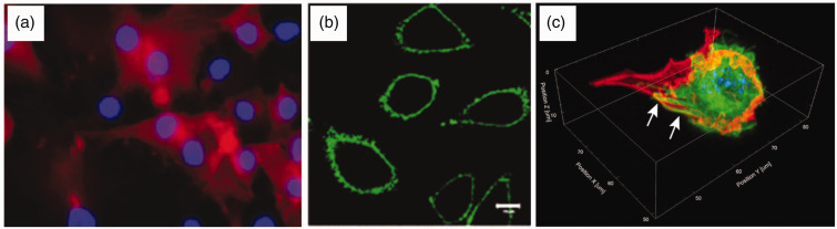
Fluorescence microscopy images of functionalized cells. (a) An example of cell
surface chemistry, with human foetal osteoblasts (nuclei-labeled blue with DAPI)
metabolically labeled with L-azidohomoalanine were conjugated to a biotinylated
alkyne that was subsequently visualized using fluorescent streptavidin (labeled red). Reprinted (adapted) with permission from Borcard et al. Bioconjugate
Chemistry 22, 1422-32 Copyright 2011 American Chemical Society. (b) An example of
non-covalent membrane labeling, in which a polyethylene glycol/oleyl chain was used
to anchor proteins such as GFP (labeled green) into NIH3T3 cells. Reproduced with kind permission from John Wiley and Sons: Kato et al. (c) An example of an extended cellular coating, whereby matrix proteins
including fibronectin (labeled red) were used to “shrink wrap” C2C12 cells
(nuclei-labeled blue with DAPI, actin fibres labeled in green and indicated with arrows). Reproduced with kind permission from Springer Science + Business Media:
Palchesko et al.,
Figure 4(e). (A color
version of this figure is available in the online journal.)
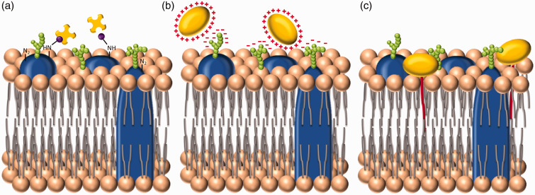
Three broad approaches to cell membrane functionalization. (a) The first method is
direct surface chemistry, performed on functional groups present on the cell
membrane. Here, for instance, amine groups present on membrane proteins have been
biotinylated (purple) to allow the addition of streptavidin (yellow). This approach
is commonly used to deliver species labeled with streptavidin or biotin. (b) The second method is to increase the cationic surface charge of the
exogenous species to facilitate attractive electrostatic interactions with
negatively charged moieties present predominantly within the glycocalyx. (c) The
third strategy uses hydrophobic interactions between a conjugated lipid tail and the
phospholipid bilayer, to anchor the exogenous species to the cell membrane. (A color
version of this figure is available in the online journal.)
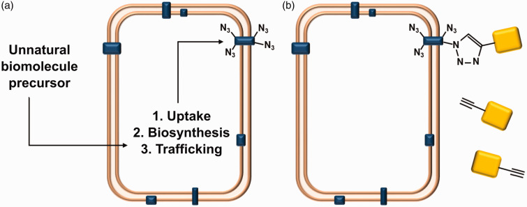
Metabolic labeling and biorthogonal chemistry. (a) Unnatural biomolecular
precursors, included as cell media additives, can be taken up by cells and become
incorporated into lipids, carbohydrates or proteins (blue), including those at the
cell membrane. (b) Metabolic labeling can be used to present reactive groups that
can bind a secondary species (yellow). This is usually mediated by orthogonal click
chemistry, in this example, an alkynated secondary species is bound to a cell
metabolically labeled with azide groups. (A color version of this figure is
available in the online journal.)
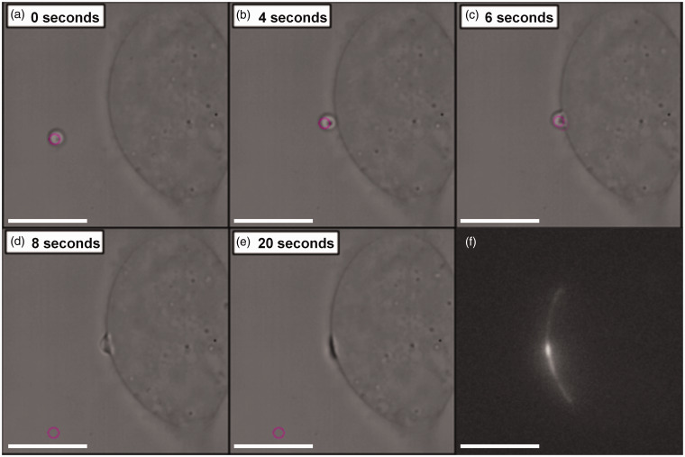
Cell paintballing using coacervate microdroplets. Armstrong et al. recently
demonstrated that membrane-free coacervate microdroplets can be actively loaded with
biomaterial payloads of protein or nucleotides, and then delivered to the cell
membrane using optical tweezers. (a)–(e) Time-lapse bright field microscope images showing an optical trap (pink
circle) maneuvering a GFP-loaded coacervate microdroplet toward a human mesenchymal
stem cell to initiate a targeted fusion event. (f) Fluorescence microscopy revealed
fluorescence emission from the GFP payload present at the site of delivery
References
-
- Derby B. Printing and prototyping of tissues and scaffolds. Science 2012; 338: 921–7. - PubMed
-
- George JC. Stem cell therapy in acute myocardial infarction: a review of clinical trials. Transl Res 2010; 155: 10–19. - PubMed
-
- Strauer BE, Kornowski R. Stem cell therapy in perspective. Circulation 2003; 107: 929–34. - PubMed
-
- Greenberg PD. Adoptive T cell therapy of tumors: mechanisms operative in the recognition and elimination of tumor cells. Adv Immunol 1991; 49: 281–355. - PubMed
Publication types
MeSH terms
Substances
LinkOut - more resources
Full Text Sources
Other Literature Sources