. 2016 Jun;7(6):434-44.
doi: 10.1007/s13238-016-0277-2. Epub 2016 May 26.

MiR-29b suppresses the proliferation and migration of osteosarcoma cells by targeting CDK6

Affiliations

MiR-29b suppresses the proliferation and migration of osteosarcoma cells by targeting CDK6

Kegan Zhu et al. Protein Cell. 2016 Jun.

Abstract

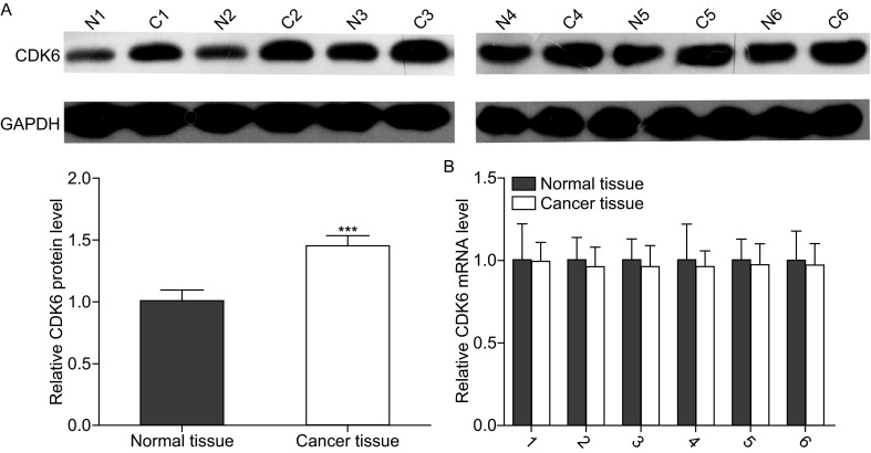
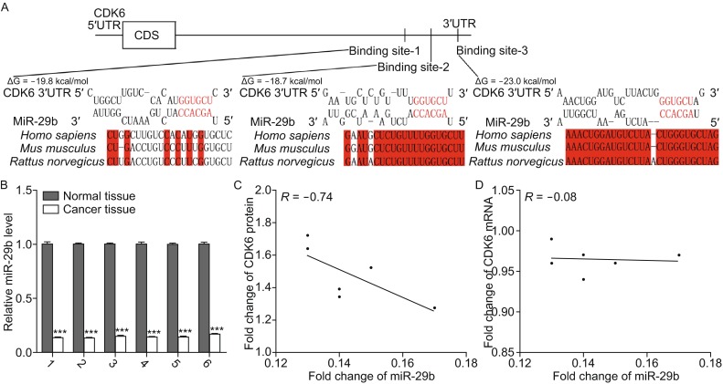
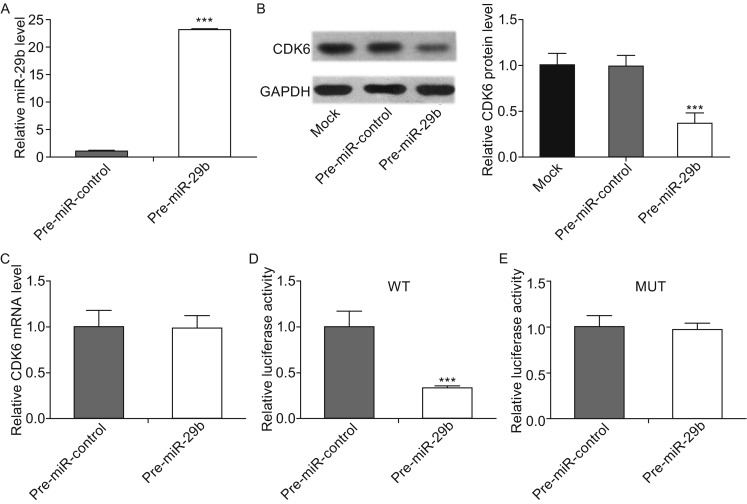
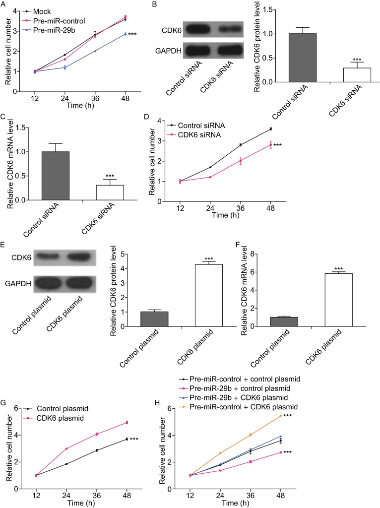
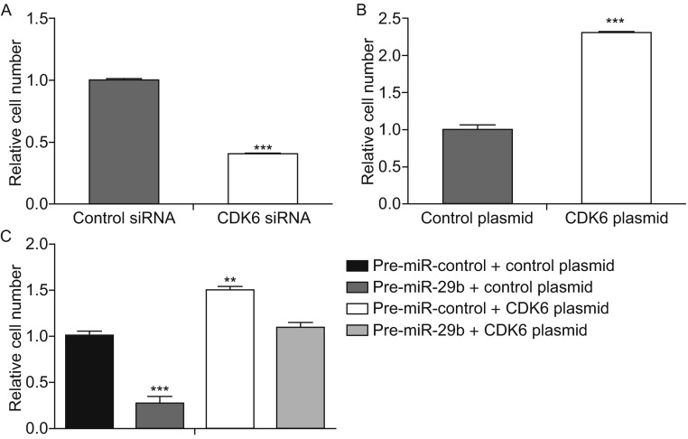
Osteosarcoma is the most common primary sarcoma of bone, and it is a leading cause of cancer death among adolescents and young adults. However, the molecular mechanism underlying osteosarcoma carcinogenesis remains poorly understood. Recently, cyclin-dependent kinase 6 (CDK6) was identified as an important oncogene. We found that CDK6 protein level, rather than CDK6 mRNA level, is much higher in osteosarcoma tissues than in normal adjacent tissues, which indicates a post-transcriptional mechanism involved in CDK6 regulation in osteosarcoma. MiRNAs are small non-coding RNAs that repress gene expression at the post-transcriptional level and have widely been shown to play important roles in many human cancers. In this study, we investigated the role of miR-29b as a novel regulator of CDK6 using bioinformatics methods. We demonstrated that CDK6 can be downregulated by miR-29b via binding to the 3'-UTR region in osteosarcoma cells. Furthermore, we identified an inverse correlation between miR-29b and CDK6 protein levels in osteosarcoma tissues. Finally, we examined the function of miR-29b-driven repression of CDK6 expression in osteosarcoma cells. The results revealed that miR-29b acts as a tumor suppressor of osteosarcoma by targeting CDK6 in the proliferation and migration processes. Taken together, our results highlight an important role for miR-29b in the regulation of CDK6 in osteosarcoma and may open new avenues for future osteosarcoma therapies.

Keywords: miR-29b; migration; osteosarcoma; proliferation; tumorigenesis.


Figures

Figure 1
Figure 1
Downregulation of CDK6 in osteosarcoma tissues. (A) Western blotting analyses of the expression levels of the CDK6 protein in 6 pairs of osteosarcoma tissues (denoted as “C”) and corresponding noncancerous tissues (denoted as “N”). Upper panel: representative image; lower panel: quantitative analysis (***P < 0.001). (B) Quantitative RT-PCR analyses of the expression levels of CDK6 mRNA in the same 6 pairs of osteosarcoma tissues and corresponding noncancerous tissues. The results were normalized to GAPDH (***P <0.001)
Figure 2
Figure 2
Inverse correlation between the miR-29b and CDK6 protein expression levels in osteosarcoma tissues. (A) Schematic description of the hypothetical duplexes formed by the interactions between the binding sites in the CDK6 3′-UTR (top) and miR-29b (bottom). The predicted free energy value of each hybrid is indicated. The seed recognition sites are denoted, and the conservation of the nucleotides in these regions across species, including human, mouse and rat, are displayed. (B) Quantitative RT-PCR analyses of the expression levels of miR-29b in the same 6 pairs of osteosarcoma tissues and corresponding noncancerous tissues. The results were normalized to U6 (***P < 0.001). (C) Pearson’s correlation scatter plot analysis of the expression levels between miR-29b and CDK6 protein in osteosarcoma tissues. (D) Pearson’s correlation scatter plot analysis of the expression levels between miR-29b and CDK6 mRNA in osteosarcoma tissues
Figure 3
Figure 3
CDK6 protein expression is inhibited by miR-29b via binding to the CDK6 3′-UTR in osteosarcoma cells. (A) Quantitative RT-PCR analyses of the expression levels of miR-29b in osteosarcoma cells after transfection with pre-miR-29b or pre-miR-control. The results were normalized to U6 (***P < 0.001). (B) Western blotting analyses of the expression levels of CDK6 protein in osteosarcoma cells after transfection with pre-miR-29b, pre-miR-control or nothing (mock). Left panel: representative images; right panel: quantitative analysis (***P < 0.001). (C) Quantitative RT-PCR analyses of the expression levels of CDK6 mRNA in the osteosarcoma cells after transfection. The results were normalized to GAPDH. (D and E) Direct recognition and combination of the CDK6 3′-UTR by miR-29b. Firefly luciferase reporters containing either wild-type (WT) or mutant (MUT) miR-29b binding sites in the CDK6 3′-UTR were co-transfected into osteosarcoma cells with either the pre-miR-control or pre-miR-29b. Twenty-four hours post-transfection, the cells were assayed using a luciferase assay kit, and the luciferase activities were normalized to the β-galactosidase levels of the control (***P < 0.001)
Figure 4
Figure 4
MiR-29b represses cell proliferation via targeting CDK6 in osteosarcoma cells. (A) The CCK8 assays were performed 12, 24, 36 and 48 h after the transfection of osteosarcoma cells with pre-miR-control, pre-miR-29b or nothing (mock). ***P < 0.001. (B) Western blotting analyses of the expression levels of the CDK6 protein in osteosarcoma cells after transfection with control siRNA or CDK6 siRNA. Left panel: representative images; right panel: quantitative analysis (***P < 0.001). (C) Quantitative RT-PCR analyses of the expression levels of CDK6 mRNA in osteosarcoma cells after transfection with control siRNA or CDK6 siRNA (***P < 0.001). (D) The CCK8 assays were performed 12, 24, 36 and 48 h after the transfection of osteosarcoma cells with control siRNA or CDK6 siRNA (***P < 0.001). (E) Western blotting analyses of the expression levels of CDK6 protein in osteosarcoma cells after transfection with control plasmid or CDK6 plasmid. Left panel: representative images; right panel: quantitative analysis (***P < 0.001). (F) Quantitative RT-PCR analyses of the expression levels of CDK6 mRNA in osteosarcoma cells after transfection with control plasmid or CDK6 plasmid (***P < 0.001). (G) The CCK8 assays were performed 12, 24, 36 and 48 h after the transfection of osteosarcoma cells with control plasmid or CDK6 plasmid (***P < 0.001). (H) The CCK8 assays were performed 12, 24, 36 and 48 h after the transfection of osteosarcoma cells with pre-miR-control plus control plasmid, pre-miR-29b plus control plasmid, pre-miR-29b plus CDK6 plasmid or pre-miR-control plus CDK6 plasmid (***P < 0.001)
Figure 5
Figure 5
MiR-29b represses cell migration via targeting CDK6 in osteosarcoma cells. (A) Transwell analyses of the migrated osteosarcoma cells after transfection with the control siRNA or CDK6 siRNA (***P < 0.001). (B) Transwell analyses of the migrated osteosarcoma cells after transfection with the control plasmid or CDK6 plasmid (***P < 0.001). (C) Transwell analyses of the migrated osteosarcoma cells after transfection with pre-miR-control plus control plasmid, pre-miR-29b plus control plasmid, pre-miR-29b plus CDK6 plasmid or pre-miR-control plus CDK6 plasmid (**P < 0.01; ***P < 0.001)

Similar articles

Cited by

References

    1. Admassi D. Osteosarcoma of medial cuniform bone. Ethiop Med J. 2009;47:305–308. - PubMed
    1. Ambros V. The functions of animal microRNAs. Nature. 2004;431:350–355. doi: 10.1038/nature02871. - DOI - PubMed
    1. Broadhead ML, Clark JCM, Myers DE, Dass CR, Choong PFM (2011) The molecular pathogenesis of osteosarcoma: a review. Sarcoma. Article No.: 959248 - PMC - PubMed
    1. Calin GA, Croce CM. MicroRNA signatures in human cancers. Nat Rev Cancer. 2006;6:857–866. doi: 10.1038/nrc1997. - DOI - PubMed
    1. Carrington JC, Ambros V. Role of microRNAs in plant and animal development. Science. 2003;301:336–338. doi: 10.1126/science.1085242. - DOI - PubMed

Publication types

MeSH terms