Review
. 2016 May 24;17 Suppl 1(Suppl 1):22.
doi: 10.1186/s12860-016-0087-7.

Molecular mechanisms regulating formation, trafficking and processing of annular gap junctions

Affiliations
Review

Molecular mechanisms regulating formation, trafficking and processing of annular gap junctions

Matthias M Falk et al. BMC Cell Biol. .

Abstract

Internalization of gap junction plaques results in the formation of annular gap junction vesicles. The factors that regulate the coordinated internalization of the gap junction plaques to form annular gap junction vesicles, and the subsequent events involved in annular gap junction processing have only relatively recently been investigated in detail. However it is becoming clear that while annular gap junction vesicles have been demonstrated to be degraded by autophagosomal and endo-lysosomal pathways, they undergo a number of additional processing events. Here, we characterize the morphology of the annular gap junction vesicle and review the current knowledge of the processes involved in their formation, fission, fusion, and degradation. In addition, we address the possibility for connexin protein recycling back to the plasma membrane to contribute to gap junction formation and intercellular communication. Information on gap junction plaque removal from the plasma membrane and the subsequent processing of annular gap junction vesicles is critical to our understanding of cell-cell communication as it relates to events regulating development, cell homeostasis, unstable proliferation of cancer cells, wound healing, changes in the ischemic heart, and many other physiological and pathological cellular phenomena.

Keywords: Annular; Clathrin; Connexin; Degradation; Dynamin; Endocytosis; Fission; Gap Junction; Lysosomes; Phosphorylation; Ubiquitination; ZO-1.


Figures
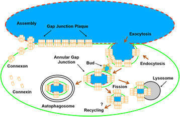
Fig. 1
Fig. 1
Schematic diagram illustrating the formation of an annular gap junction from a gap junction plaque and its subsequent processing. Newly synthesized connexin (Cx) proteins assemble into a six-protein oligomer called a connexon. Connexons are then transported to and inserted into the plasma membrane. During cell-cell contact, a hemichannel can dock head-on with a hemichannel from an apposing cell and cluster to form a gap junction plaque. Once the plaque is no longer needed for cell-cell communication, or a cell becomes migratory, a portion of the plaque (usually the central portion) or the entire plaque is internalized to form an annular gap junction. The annular gap junction may then be degraded via a number of processes, including endo-lysosomal, autophagosomal, or a fission process followed by lysosomal degradation (as shown). It has been suggested that annular gap junction vesicles or free connexin proteins in cytoplasmic membranes that are released during annular gap junction vesicle degradation may recycle back to the cell surface to participate in the formation of new, or the addition to existing gap junction plaques. Black arrows depict structural components, while maroon arrows depict cellular processes
Fig. 2
Fig. 2
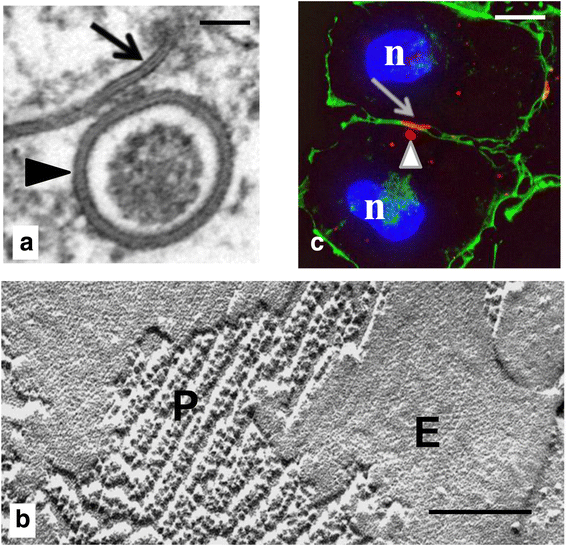
Gap junction plaques (arrows) and annular gap junction vesicles (arrowheads) shown with transmission electron (a), freeze fracture electron (b), and immunofluorescence microscopy (c). Staining cortical actin (green in c) helps to define the cell borders. The protoplasmic (P) and extracellular (E) fracture faces have been labeled in the replica of the gap junction plaque (in b). Nucleus = n. Bars: 100 nm in (a), 60 nm in (b), and 10 μm in (c). (a from ref. [58] and b from ref. [206])
Fig. 3
Fig. 3
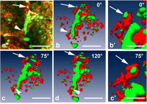
Immunocytochemistry (a) and 3D-volume reconstructions (b-d) demonstrating in detail the association of clathrin (red) with a Cx43 (green) gap junction plaque. An area of the gap junction plaque can be appreciated in the rotated views as a bud that likely has not yet detached from the plaque (a-d arrowheads). It can be confirmed in the rotated views that an annular gap junction is intimately associated with clathrin (arrows a-d). A rotation around the Y-axis in the 3D-reconstruction images allows more information to be obtained on the morphology of the gap junction structures as well as their relationship to clathrin. The images seen in (b) and (c) (arrows) have been enlarged (b'-c'). The 3D-reconstruction was rendered with the Amira 3D Software for Life Sciences, FEI™ (Hillsboro, Oregon). Bar: 5 μm in (a-d), 1.5 μm in (b’, c’)
Fig. 4
Fig. 4
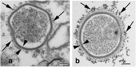
Quantum dot immuno-electron microscopy demonstrating annular gap junction vesicles decorated with phosphorylated Cx43 (a) and clathrin (b) (arrows). Note the characteristic annular gap junction double-membrane, which helps to distinguish the annular gap junction vesicle from other membraned cellular structures (see arrowheads). Bars: 100 nm
Fig. 5
Fig. 5
Internalization of a gap junction plaque. a Schematic depicting the internalization of a complete gap junction. The process leads to the formation of an annular gap junction (AGJ) vesicle in the cytoplasm of one of the coupled cells. b, c The process schemed in (a) imaged by time-lapse microscopy in Cx43-GFP expressing HeLa cells (only fluorescence shown in (b), merged fluorescence and DIC channels shown in (c)). Note that the small gap junction plaque in (b) (depicted by the upper arrow) does not internalize and remains in the plasma membrane while the large gap junction (depicted by the lower arrow) invaginates into the left cell of the coupled pair and forms an annular gap junction; and that in (c) a new gap junction (depicted with arrow) forms at the location were the previous internalized gap junction plaque (depicted with arrowhead) was localized. Time in c is in hours:minutes. (B.N.G. Giepmans and C. Lehmann recorded time-lapse movie sequences shown in (b) and (c), respectively when working in the Falk lab.)
Fig. 6
Fig. 6
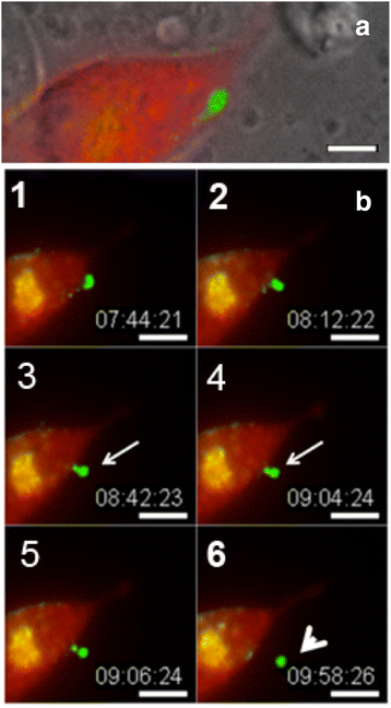
Time-lapse image sequence of cells expressing Cx43-GFP. As evident from the phase/fluorescence overlay image (a) one of the two contacting cells is expressing RFP-tagged clathrin (red). In the corresponding montage of these cells, the gap junction plaque invaginates to form a gap junction bud (arrow), which is then scissored from the membrane to produce an annular gap junction vesicle (arrowhead) (b, 1-6). Time is in hours:minutes:seconds. Bar: 15 μm. (From ref. [58])
Fig. 7
Fig. 7
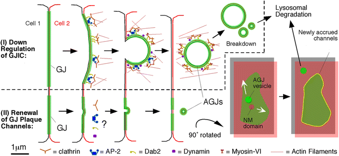
Schematic representation of the pathways that lead to the internalization of entire gap junction plaques (I) and of central plaque portions (II), annular gap junction formation, fission, and degradation. Whether clathrin and clathrin-accessory proteins are involved in the internalization of small gap junction vesicles shown in (II) has not been determined, however is likely based on our EM analyses. Accrual of new channels (yellow line circumscribing the green gap junction plaque) is accompanied by the simultaneous internalization of central plaque portions consistent with previously published observations [63, 72]. Clathrin and accessory proteins are shown in patches in accordance to the appearance of clathrin on gap junction plaques [61], and the current thinking that clathrin may provide a scaffold for directed actin assembly, facilitating internalization of large structures such as gap junctions, viruses, and pathogenic bacteria [207, 208]. The manner in which clathrin and accessory proteins are drawn still remains somewhat speculative. NM = Connexin-free junctional membrane domain. (From ref. [73])
Fig. 8
Fig. 8
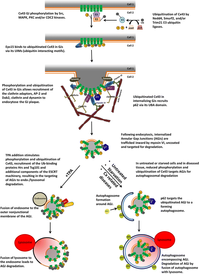
Schematic representation of the signals and players that participate in the steps that lead to gap junction internalization, formation of annular gap junctions in the cytoplasm of the acceptor cell, and annular gap junction degradation through the phago-lysosomal (bottom right) or the endo-lysosomal (bottom left) pathway based on published studies. Abbreviations are: AGJ, annular gap junction; CLASPs, clathrin-associated sorting proteins; ESCRT, endosomal sorting complexes required for transport; GJ, gap junction; p62, sequestosome 1/SQSTM1; UBA, ubiquitin-associated domain; UIMs, ubiquitin-interacting motifs. (From ref. [182])
Fig. 9
Fig. 9
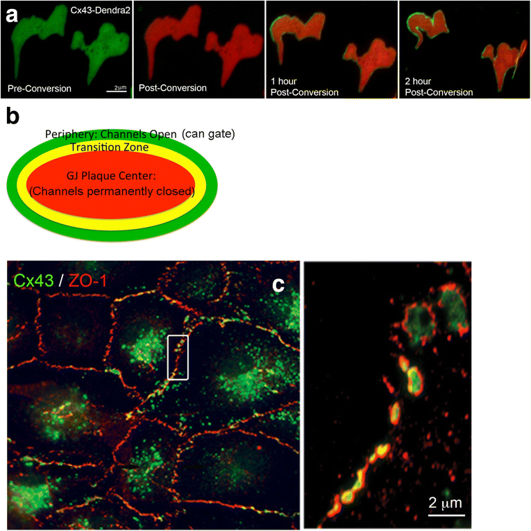
Gap junction plaque assembly and structure. a Photoconversion of Cx43-Dendra2 reveals accrual of newly synthesized gap junction channels (non-converted, green) along the outer edge of gap junction plaques (permanently photoconverted from green to red; shown for two gap junction plaques viewed en face/onto the plaque surface). (From ref. [73].) b Schematic model of a gap junction depicting our hypothesized plaque organization. c Cx43 gap junctions (green) colocalizing with the scaffolding protein, ZO-1 (red), generating a typical staining along the rim of gap junction plaques [130], shown in fixed endogenously Cx43 expressing primary pulmonary artery endothelial cells (PAECs). The boxed area is shown enlarged on the right. (From ref. [131])
Fig. 10
Fig. 10
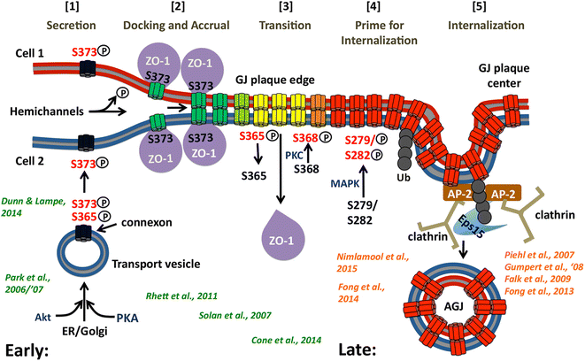
Scheme depicting the molecular signals in the Cx43-C-terminal domain hypothesized to regulate gap junction assembly and internalization based on our own (orange) and colleagues’ (green) findings. Steps [1 – 5] trigger and coordinate the transition from functional (green) into non-functional, internalization-prone gap junction channels (yellow, orange) that then are primed via post-translational modifications to allow interaction with clathrin components to mediate their internalization (red). The color scheme of the channels corresponds to the schematic gap junction shown in Fig. 9b
Fig. 11
Fig. 11
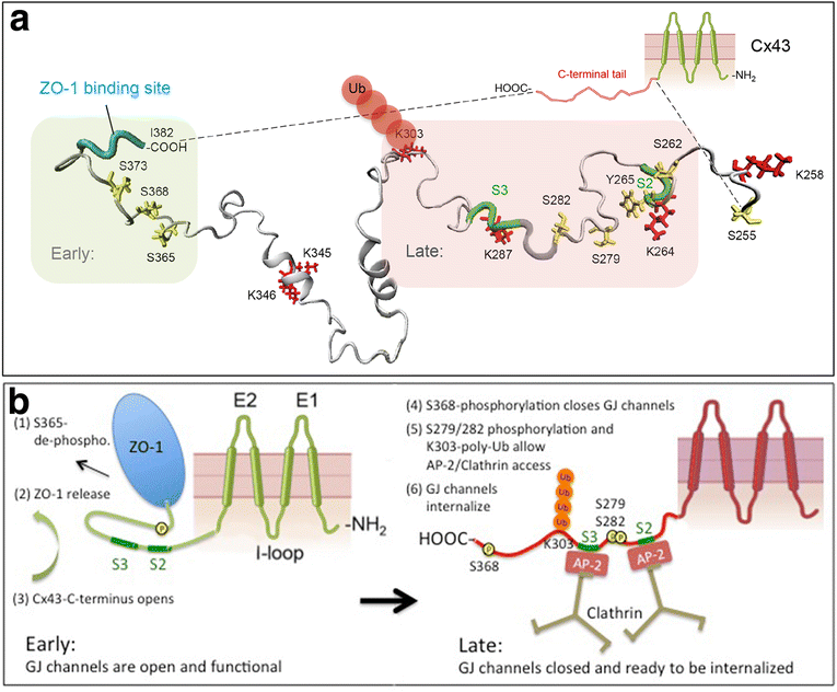
Scheme depicting how access of clathrin to Cx43 might be regulated. a Interestingly, all proposed Cx43 modifications relevant to Cx43 gap junction internalization cluster into two domains, ‘early’ occurring on residues located juxtaposed to the C-terminal ZO-1 binding site (shaded green), and ‘late’ occurring on residues located juxtaposed to the S2, S3 AP-2 (Eps 15)/clathrin binding sites (shaded red). The lowest energy 3D solution NMR structure of the Cx43-CT revealing the location of critical residues solved by Sorgen and colleagues [117] is shown. b We propose that a conformational change of the Cx43-C-terminal domain (CT) triggered by serine 365 de-phosphorylation [116] opens up the Cx43-CT allowing MAPK to access and phosphorylate S279/282 (and eventually also S262 and S255); and E3-ubiquitin ligases to bind to and ubiquitinate Cx43 (presumably on lysine 303) to promote AP-2 (and/or Eps15) to access the YXXΦ -binding motifs (S2, 265YAYF268; S3, 286YKLV289), recruit clathrin and internalize gap junctions/central gap junction plaque portions
Fig. 12
Fig. 12
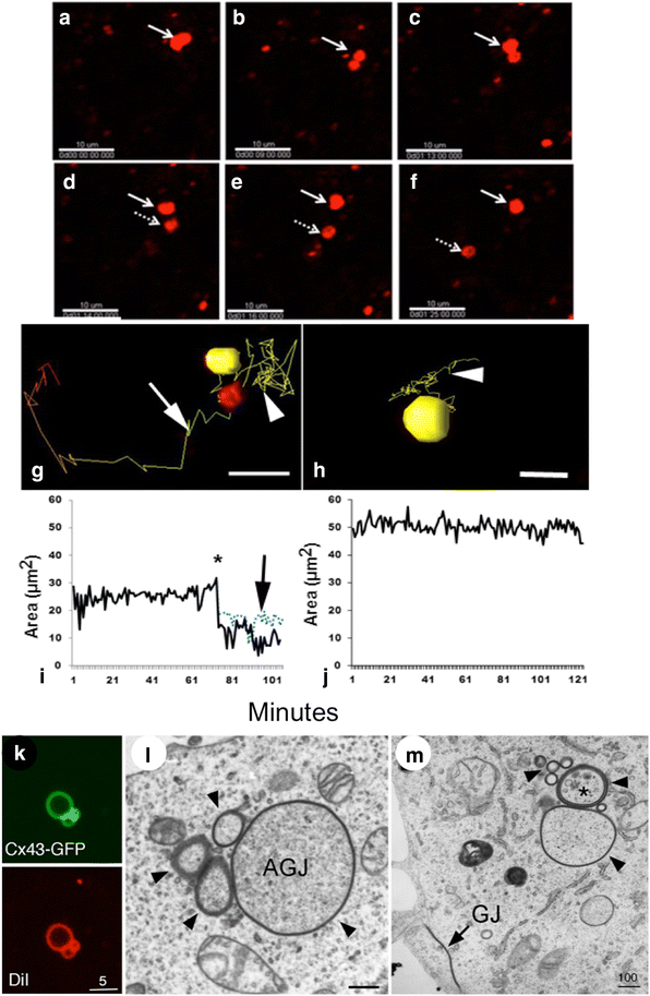
Annular gap junction vesicle fission. In the time-lapse imaging montage the splitting of an annular gap junction vesicle is demonstrated (a-f). Once the split occurs, the two annular gap junction vesicles (arrow and dashed arrow) move away from one another (d-f). The path and size of annular gap junction vesicles is depicted in the animated 3D reconstruction time-lapse tracking (g, h) and in the corresponding graphs (i, j). Note the yellow line depicting the annular gap junction movement path before (arrowhead) and after the split (arrow) (g) and the corresponding changes in size after fission (i). Inhibiting dynamin function blocks annular gap junction splitting (h, j). Clusters of annular gap junction vesicles, which would be consistent with the splitting/budding process, can be seen with fluorescence light microscopy in (k), and transmission electron microscopy (AGJ, marked with arrowheads in l, m) in the cytoplasm. Note the gap junction plaque (GJ) in the plasma membranes in (m). Membranes have been labeled with DiI (red in k). Bars: 10 μm (a-f), 5 μm (g, h, k), 100 nm (l, m). (a-j from ref. [58], and k-m from ref. [61])

Similar articles

Cited by

References

    1. Goodenough DA, Goliger JA, Paul DL. Connexins, Connexons, and intercellular communication (Review) Ann Rev Biochem. 1996;65:475–502. doi: 10.1146/annurev.bi.65.070196.002355. - DOI - PubMed
    1. Kumar NM, Gilula NB. Molecular biology and genetics of gap junction channels. Semin Cell Biol. 1992;3(1):3–16. doi: 10.1016/S1043-4682(10)80003-0. - DOI - PubMed
    1. Loewenstein WR, Rose B. The cell-cell channel in the control of growth. Semin Cell Biol. 1992;3(1):59–79. doi: 10.1016/S1043-4682(10)80008-X. - DOI - PubMed
    1. Wei CJ, Xu X, Lo CW. Connexins and cell signaling in development and disease. Annu Rev Cell Dev Biol. 2004;20:812–38. doi: 10.1146/annurev.cellbio.19.111301.144309. - DOI - PubMed
    1. Grek CL, Rhett JM, Ghatnekar GS. Cardiac to cancer: connecting connexins to clinical opportunity. FEBS Lett. 2014;588(8):1349–64. doi: 10.1016/j.febslet.2014.02.047. - DOI - PMC - PubMed

LinkOut - more resources