

JNK1 ablation in mice confers long-term metabolic protection from diet-induced obesity at the cost of moderate skin oxidative damage
- PMID: 27230858
- PMCID: PMC6191003
- DOI: 10.1096/fj.201600393R
JNK1 ablation in mice confers long-term metabolic protection from diet-induced obesity at the cost of moderate skin oxidative damage
Abstract
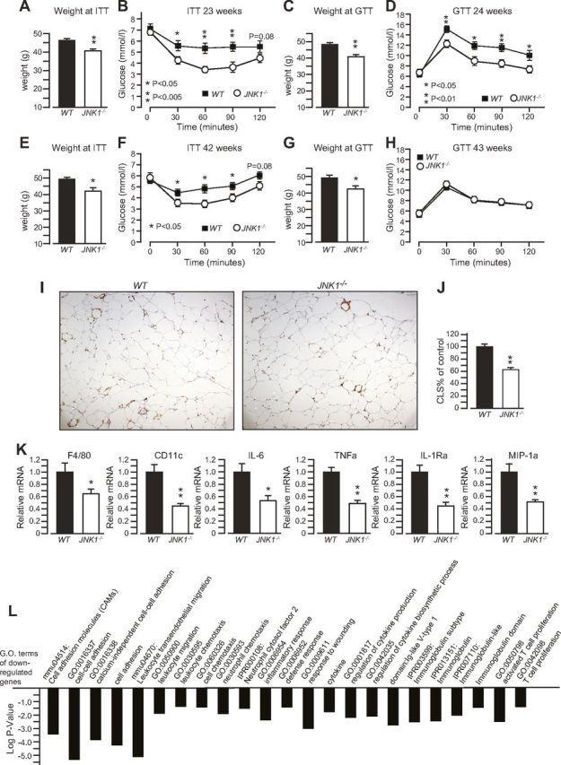
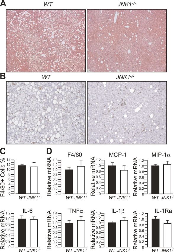
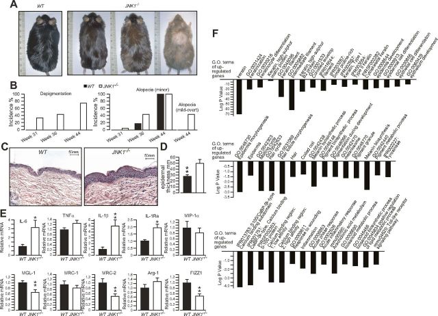
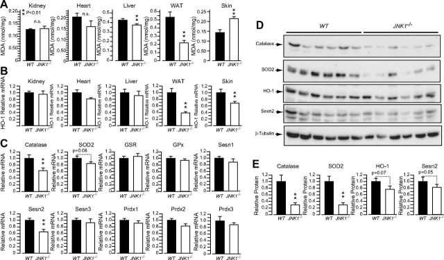
Obesity and insulin resistance are associated with oxidative stress, which may be implicated in the progression of obesity-related diseases. The kinase JNK1 has emerged as a promising drug target for the treatment of obesity and type 2 diabetes. JNK1 is also a key mediator of the oxidative stress response, which can promote cell death or survival, depending on the magnitude and context of its activation. In this article, we describe a study in which the long-term effects of JNK1 inactivation on glucose homeostasis and oxidative stress in obese mice were investigated for the first time. Mice lacking JNK1 (JNK1(-/-)) were fed an obesogenic high-fat diet (HFD) for a long period. JNK1(-/-) mice fed an HFD for the long term had reduced expression of antioxidant genes in their skin, more skin oxidative damage, and increased epidermal thickness and inflammation compared with the effects in control wild-type mice. However, we also observed that the protection from obesity, adipose tissue inflammation, steatosis, and insulin resistance, conferred by JNK1 ablation, was sustained over a long period and was paralleled by decreased oxidative damage in fat and liver. We conclude that compounds targeting JNK1 activity in brain and adipose tissue, which do not accumulate in the skin, may be safer and most effective.-Becattini, B., Zani, F., Breasson, L., Sardi, C., D'Agostino, V. G., Choo, M.-K., Provenzani, A., Park, J. M., Solinas, G. JNK1 ablation in mice confers long-term metabolic protection from diet-induced obesity at the cost of moderate skin oxidative damage.
Keywords: antioxidants; insulin resistance; metabolic inflammation; oxidative stress tolerance; type-2 diabetes.
© FASEB.
Conflict of interest statement
This work was supported by grants from the Swiss National Science Foundation, the Swedish Diabetes Foundation, the Swedish Research Council, and a startup package from the University of Gothenburg (to G.S.). Author contributions: B. Becattini and F. Zani performed most of the experiments, analyzed data, and contributed to the discussion; L. Breasson and C. Sardi performed experiments; V. G. D’Agostino and A. Provenzani performed the DNA microarray analysis; M.-K. Choo and J. M. Park performed the skin histopathological analysis and discussed the skin data; G. Solinas conceived the project, the experimental design, and wrote the manuscript; B. Becattini, F. Zani, A. Provenzani, M.-K. Choo and J. M. Park, significantly contributed to the writing of the manuscript with their comments; and all authors approved the paper. The authors declare no conflicts of interest.
Figures

Similar articles
-
Role of muscle c-Jun NH2-terminal kinase 1 in obesity-induced insulin resistance.Mol Cell Biol. 2010 Jan;30(1):106-15. doi: 10.1128/MCB.01162-09. Mol Cell Biol. 2010. PMID: 19841069 Free PMC article.
-
PI3Kγ ablation does not promote diabetes in db/db mice, but improves insulin sensitivity and reduces pancreatic β-cell apoptosis.FASEB J. 2018 Jan;32(1):319-329. doi: 10.1096/fj.201700372RR. Epub 2017 Sep 13. FASEB J. 2018. PMID: 28904022
-
Differential effects of JNK1 and JNK2 inhibition on murine steatohepatitis and insulin resistance.Hepatology. 2009 Jan;49(1):87-96. doi: 10.1002/hep.22578. Hepatology. 2009. PMID: 19053047 Free PMC article.
-
cJun NH2-terminal kinase 1 (JNK1): roles in metabolic regulation of insulin resistance.Trends Biochem Sci. 2010 Sep;35(9):490-6. doi: 10.1016/j.tibs.2010.04.004. Epub 2010 May 7. Trends Biochem Sci. 2010. PMID: 20452774 Free PMC article. Review.
-
Interplay between diet-induced obesity and oxidative stress: Comparison between Drosophila and mammals.Comp Biochem Physiol A Mol Integr Physiol. 2019 Feb;228:18-28. doi: 10.1016/j.cbpa.2018.09.027. Epub 2018 Oct 30. Comp Biochem Physiol A Mol Integr Physiol. 2019. PMID: 30385171 Review.
Cited by
-
JNK at the crossroad of obesity, insulin resistance, and cell stress response.Mol Metab. 2016 Dec 8;6(2):174-184. doi: 10.1016/j.molmet.2016.12.001. eCollection 2017 Feb. Mol Metab. 2016. PMID: 28180059 Free PMC article. Review.
-
Long-term, high-fat feeding exacerbates short-term increases in adipose mitochondrial reactive oxygen species, without impairing mitochondrial respiration.Am J Physiol Endocrinol Metab. 2020 Aug 1;319(2):E376-E387. doi: 10.1152/ajpendo.00028.2020. Epub 2020 Jun 16. Am J Physiol Endocrinol Metab. 2020. PMID: 32543945 Free PMC article.
-
Antioxidant, antihyperglycemic, and antidiabetic activity of Apis mellifera bee tea.PLoS One. 2018 Jun 5;13(6):e0197071. doi: 10.1371/journal.pone.0197071. eCollection 2018. PLoS One. 2018. PMID: 29870561 Free PMC article.
-
JNK1 and JNK3: divergent functions in hippocampal metabolic-cognitive function.Mol Med. 2022 May 4;28(1):48. doi: 10.1186/s10020-022-00471-y. Mol Med. 2022. PMID: 35508978 Free PMC article.
-
c-Jun N-terminal Kinase 1 ablation protects against metabolic-induced hippocampal cognitive impairments.J Mol Med (Berl). 2019 Dec;97(12):1723-1733. doi: 10.1007/s00109-019-01856-z. Epub 2019 Dec 3. J Mol Med (Berl). 2019. PMID: 31797011
References
-
- Solinas G. (2012) Molecular pathways linking metabolic inflammation and thermogenesis. Obes. Rev. (Suppl 2), 69–82 - PubMed
-
- Solinas G., Karin M. (2010) JNK1 and IKKbeta: molecular links between obesity and metabolic dysfunction. FASEB J. , 2596–2611 - PubMed
-
- Unger R. H., Clark G. O., Scherer P. E., Orci L. (2010) Lipid homeostasis, lipotoxicity and the metabolic syndrome. Biochim. Biophys. Acta , 209–214 - PubMed
-
- Hotamisligil G. S. (2006) Inflammation and metabolic disorders. Nature , 860–867 - PubMed
Publication types
MeSH terms
Substances
Grants and funding
LinkOut - more resources
Full Text Sources
Other Literature Sources
Medical
Molecular Biology Databases
Research Materials
Miscellaneous
