Randomized Controlled Trial
. 2016 Jun 2;98(6):1208-1219.
doi: 10.1016/j.ajhg.2016.05.014. Epub 2016 May 23.

Mosaic Loss of Chromosome Y in Blood Is Associated with Alzheimer Disease

Affiliations
Randomized Controlled Trial

Mosaic Loss of Chromosome Y in Blood Is Associated with Alzheimer Disease

Jan P Dumanski et al. Am J Hum Genet. .

Abstract

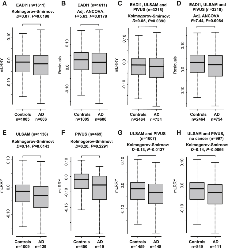
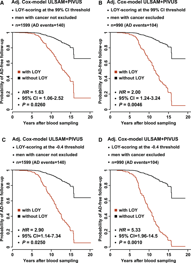
Men have a shorter life expectancy compared with women but the underlying factor(s) are not clear. Late-onset, sporadic Alzheimer disease (AD) is a common and lethal neurodegenerative disorder and many germline inherited variants have been found to influence the risk of developing AD. Our previous results show that a fundamentally different genetic variant, i.e., lifetime-acquired loss of chromosome Y (LOY) in blood cells, is associated with all-cause mortality and an increased risk of non-hematological tumors and that LOY could be induced by tobacco smoking. We tested here a hypothesis that men with LOY are more susceptible to AD and show that LOY is associated with AD in three independent studies of different types. In a case-control study, males with AD diagnosis had higher degree of LOY mosaicism (adjusted odds ratio = 2.80, p = 0.0184, AD events = 606). Furthermore, in two prospective studies, men with LOY at blood sampling had greater risk for incident AD diagnosis during follow-up time (hazard ratio [HR] = 6.80, 95% confidence interval [95% CI] = 2.16-21.43, AD events = 140, p = 0.0011). Thus, LOY in blood is associated with risks of both AD and cancer, suggesting a role of LOY in blood cells on disease processes in other tissues, possibly via defective immunosurveillance. As a male-specific risk factor, LOY might explain why males on average live shorter lives than females.


Figures

Figure 1
Figure 1
Reproducible Estimations of LOY via Two Independent Technologies Validation of LOY mosaicism detected by SNP-array data by whole-genome next-generation sequencing (WGS) in 183 EADI1 participants. This was performed by estimating ploidy values from about 30× NGS data using the FREEC software.
Figure 2
Figure 2
Mosaic LOY in Blood Cells Increases with Participant Sampling Age (A) Illustration of the association between of LOY and age of sampling in all 3,218 subjects from three independent cohorts included in the analyses. Linear regression shows that LOY (i.e., mLRRY) was associated with sampling age (ANOVA; F(1,3216) = 26.79, p < 0.0001). (B–D) Corresponding plots of the data from ULSAM (B), PIVUS (C), and EADI1 (D) cohorts. The dotted horizontal lines show the cut-off used for LOY scoring at the 99% confidence interval in the three independent cohorts. (E) The increasing degree of LOY in four different age groups in the three included studies and p values adjusted for multiple testing using Tukey’s method is shown. The whiskers extend to illustrate the 1.5 inter-quantile range of the total variation in each age group. (F) Summary of the observed frequencies of LOY in the different age groups plotted in (E).
Figure 3
Figure 3
Men Diagnosed with Alzheimer Disease Had on Average a Higher Level of LOY in Blood Cells Compared to AD-free Controls (A and B) Unadjusted and adjusted analyses performed in the case-control EADI1 study (n = 1,611). Men with AD had significantly higher level of LOY compared to controls (A, unadjusted Kolmogorov-Smirnov test: D = 0.07, p = 0.0198). Difference in LOY between subjects with AD and controls by plotting the adjusted residuals from an ANCOVA model fitting the effects from sampling age and APOE genotype (B). Also in this model subjects with AD diagnosis had a significantly higher level of LOY in blood compared to the control subjects (ANCOVA: F(1,1604) = 5.63, p = 0.0178). (C and D) Results from analogous comparisons after pooling data from three independent cohorts, i.e., EADI1, ULSAM, and PIVUS (n = 3,218) and a significant difference in mLRRY values between all men with AD diagnosis (prevalent and incident) compared to control subjects in unadjusted (C, Kolmogorov-Smirnov test: D = 0.05, p = 0.0390) as well as adjusted (D, ANCOVA: F(1,3085) = 7.44, p = 0.0064) tests. In (D) we plotted the adjusted residuals using the same method as described for (B). (E–H) Levels of LOY observed in men with and without AD diagnosis (prevalent and incident) in the ULSAM (E) and PIVUS (F) cohorts separately as well as pooled together (G) and after removing subjects diagnosed with cancer (H). The whiskers in all boxplots extend to illustrate the 1.5 inter-quantile range of the total variation in each group.
Figure 4
Figure 4
Cox Proportional Hazards Regression Models Adjusting for Potential Confounders Show that Men with Mosaic LOY at Blood Cells at Time for Sampling Were More Likely to Be Diagnosed with AD during the Follow-up Time Probabilities for AD-free follow-up time are illustrated using red and black curves for men with and without LOY, respectively. Participants were scored as with or without LOY using two defined thresholds. Participants with an mLRRY value lower than the 99% confidence limit of the experimental variation were scored with LOY (A and B) and a threshold at mLRRY ≤ −0.4 was used (C and D). The analyses were performed using pooled data from the ULSAM and PIVUS studies. Shown are analyses with all men (A and C; n = 1,599, AD events = 140) and analogous analyses after excluding men with any cancer (B and D; n = 990, AD events = 104). The effects from all confounders in the models are given in Tables S4, S5, S7, and S8.

References

    1. Bettens K., Sleegers K., Van Broeckhoven C. Genetic insights in Alzheimer’s disease. Lancet Neurol. 2013;12:92–104. - PubMed
    1. Hardy J., Selkoe D.J. The amyloid hypothesis of Alzheimer’s disease: progress and problems on the road to therapeutics. Science. 2002;297:353–356. - PubMed
    1. Gatz M., Reynolds C.A., Fratiglioni L., Johansson B., Mortimer J.A., Berg S., Fiske A., Pedersen N.L. Role of genes and environments for explaining Alzheimer disease. Arch. Gen. Psychiatry. 2006;63:168–174. - PubMed
    1. Genin E., Hannequin D., Wallon D., Sleegers K., Hiltunen M., Combarros O., Bullido M.J., Engelborghs S., De Deyn P., Berr C. APOE and Alzheimer disease: a major gene with semi-dominant inheritance. Mol. Psychiatry. 2011;16:903–907. - PMC - PubMed
    1. Lambert J.C., Ibrahim-Verbaas C.A., Harold D., Naj A.C., Sims R., Bellenguez C., DeStafano A.L., Bis J.C., Beecham G.W., Grenier-Boley B., European Alzheimer’s Disease Initiative (EADI) Genetic and Environmental Risk in Alzheimer’s Disease. Alzheimer’s Disease Genetic Consortium. Cohorts for Heart and Aging Research in Genomic Epidemiology Meta-analysis of 74,046 individuals identifies 11 new susceptibility loci for Alzheimer’s disease. Nat. Genet. 2013;45:1452–1458. - PMC - PubMed

Publication types