

[Villonodular synovitis of the ankle, an uncommon location: a case report]
- PMID: 27231502
- PMCID: PMC4867721
- DOI: 10.11604/pamj.2016.23.90.8838
[Villonodular synovitis of the ankle, an uncommon location: a case report]
Abstract
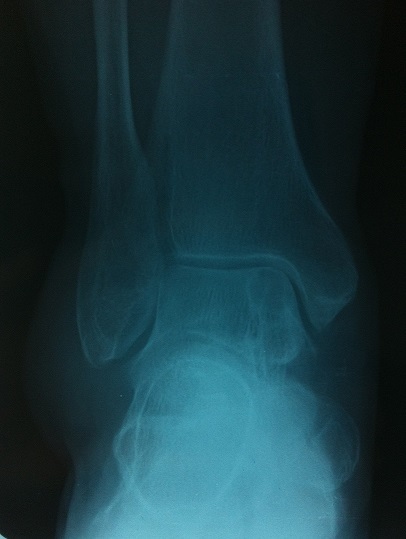
Pigmented villonodular synovitis (PVNS) is a rare pseudotumoral proliferative benign condition of unknown etiology, affecting synovial joints. It can also develop in the synovial bursae and in the tendon sheaths. It usually affects large joints, such as the knee and the hip. Ankle is a rare location, with only a few cases reported in the literature. We report a case of a 52 year old patient with a PVS of the right ankle. She received a subtotal synovectomy. After 2 years of follow-up there was no clinical recurrence.
La synovite villonodulaire (SVN) est une prolifération pseudotumorale bénigne rare de la synoviale articulaire, d’étiologie inconnue. Elle peut aussi se développer au sein des bourses séreuses, des gaines tendineuses. Généralement, elle atteint les grosses articulations notamment le genou et la hanche. La localisation de la cheville est rare, avec seulement quelques cas publiés dans la littérature. Nous rapportons un cas de patiente de 52 ans présentant une SVN de la cheville droite. Elle a bénéficié d'une synovectomie subtotale. A deux ans de recul, il n'y avait pas de récidive clinique.
Keywords: Villonodular synovitis; ankle; synovectomy.
Figures


References
-
- Tekaya R, Haouel M, Kammoun SH, et al. Synovite villonodulaire de l'articulation sub-talaire. Médecine et Chirurgie du Pied. 2011;27(4):106–108.
-
- Jaffe HL, Lichtenstein L, Sutro CJ. Pigmented villonodularsynovitis,bursitis and tenosynovitis. Archives Pathology. 1941;31(3):731–765.
-
- Myers BW, Masi AT. Pigmented villonodular synovitis and tenosynovitis: a clinical epidemiologic study of 166 cases and literature review. Medicine. 1980;59(3):223–238. - PubMed
-
- Sharma H, Rana B, Mahendra A, et al. Outcome of 17 pigmented villonodular synovitis of the knee at 6 years mean follow-up. Knee. 2007;14(5):390–4. - PubMed
-
- Murphey MD, Rhee JH, Lewis RB, et al. Pigmented villonodular synovitis: radiologic-pathologic correlation. Radiographics. 2008;28(5):1493–518. - PubMed
Publication types
MeSH terms
LinkOut - more resources
Full Text Sources
Other Literature Sources
Research Materials
