. 2016 Feb 19;7(3):943-8.
doi: 10.1364/BOE.7.000943. eCollection 2016 Mar 1.

Optically trapping tumor cells to assess differentiation and prognosis of cancers

Affiliations

Optically trapping tumor cells to assess differentiation and prognosis of cancers

M Pradhan et al. Biomed Opt Express. .

Abstract

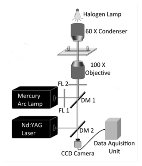
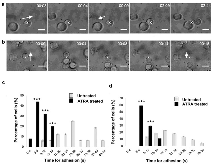
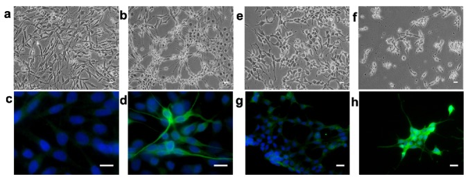
We report an optical trapping method that may enable assessment of the differentiation status of cancerous cells by determining the minimum time required for cell-cell adhesion to occur. A single, live cell is trapped and brought into close proximity of another; the minimum contact time required for cell-cell adhesion to occur is measured using transformed cells from neural tumor cell lines: the human neuroblastoma SK-N-SH and rat C6 glioma cells. Earlier work on live adult rat hippocampal neural progenitors/stem cells had shown that a contact minimum of ~5 s was required for cells to adhere to each other. We now find the average minimum time for adhesion of cells from both tumor cell lines to substantially increase to ~20-25 s, in some cases up to 45 s. Upon in vitro differentiation of these cells with all-trans retinoic acid the average minimum time reverts to ~5-7 s. This proof-of-concept study indicates that optical trapping may be a quick, sensitive, and specific method for determining differentiation status and, thereby, the prognosis of cancer cells.

Keywords: (000.1430) Biology and medicine; (170.1530) Cell analysis.


Figures

Fig. 1
Fig. 1
Schematic diagram of our optical trap (see text). Cell trapping occurred on the microscope slide placed after the oil-immersed 100X objective. DM: dichroic mirrors, FL: filters.
Fig. 2
Fig. 2
Dependence of minimum time for cell-cell adhesion on differentiation status of malignant cells. Time lapse images from real time movies depicting (a) an untreated C6 glioma cell made to approach another (Visualization 1). The trap is moved after every (n + 5) second where n = 0, 5, 10, 15 s. Cells were allowed to remain in contact till they could not be pulled apart by the trap: the time taken was designated the minimum time for cell-cell adhesion. The white cross denotes the trapped cell. In the first three frames, another cell is made to approach the trapped cell in the direction of the arrow. As the trap is moved away after 9 s, the cells are seen to separate. Eventually, the cells remain adhered to each other (last two frames). (b) Similar time lapse images from real-time movies for ATRA-treated C6 glioma cells showing adherence after 4 s contact (Visualization 2). (c), (d) Percentage of undifferentiated and ATRA-treated cells undergoing irreversible cell adhesion at different time points (see text). (c) SK-N-SH cells; (d) C6 glioma cells. *** denotes statistical significance where p<0.001 for adhesion times of untreated and ATRA-treated cells.
Fig. 3
Fig. 3
Bright-field microscopy images of (a) untreated and (b) ATRA-treated C6 glioma cells, and of (e) untreated and (f) ATRA-treated SK-N-SH neuroblastoma cells. Expression of GFAP in untreated C6 cells (c) and in ATRA-treated C6 cells (d). Expression of β-tubulin III in undifferentiated SK-N-SH cells (g) showing low expression in a few cells and in ATRA-treated SK-N-SH cells (h) with moderate or intense expression in most cells. The white scale bar denotes 10 µm.

References

    1. Kornyshev A. A., “From biologically-inspired physics to physics-inspired biology,” J. Phys. Condens. Matter 22(41), 410401 (2010).10.1088/0953-8984/22/41/410401 - DOI - PubMed
    1. Gruebele M., Thirumalai D., “Perspective: Reaches of chemical physics in biology,” J. Chem. Phys. 139(12), 121701 (2013).10.1063/1.4820139 - DOI - PMC - PubMed
    1. Mathur D., “Biology-inspired AMO physics,” J. Phys. B 48(2), 022001 (2015).10.1088/0953-4075/48/2/022001 - DOI
    1. Sung Y., Lue N., Hamza B., Martel J., Irimia D., Dasari R. R., Choi W., Yaqoob Z., So P., “Three-dimensional holographic refractive-index measurement of continuously flowing cells in a microfluidic channel,” Phys. Rev. Appl. 1(1), 014002 (2014).10.1103/PhysRevApplied.1.014002 - DOI - PMC - PubMed
    1. Cross S. E., Jin Y. S., Rao J., Gimzewski J. K., “Nanomechanical analysis of cells from cancer patients,” Nat. Nanotechnol. 2(12), 780–783 (2007).10.1038/nnano.2007.388 - DOI - PubMed

LinkOut - more resources