. 2016 Sep:7:1-9.
doi: 10.1016/j.bbrep.2016.05.009.

High salt induces anti-inflammatory MΦ2-like phenotype in peripheral macrophages

Affiliations

High salt induces anti-inflammatory MΦ2-like phenotype in peripheral macrophages

Suneetha Amara et al. Biochem Biophys Rep. 2016 Sep.

Abstract

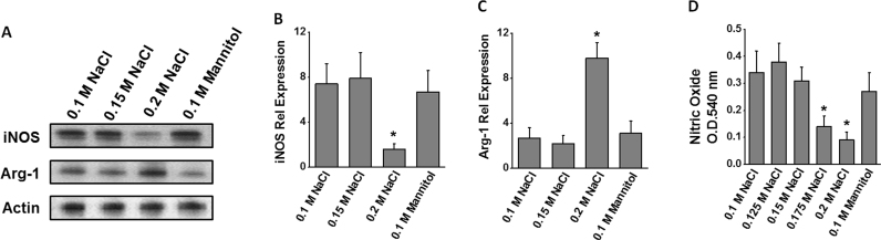
Macrophages play a critical role in inflammation and antigen-presentation. Abnormal macrophage function has been attributed in autoimmune diseases and cancer progression. Recent evidence suggests that high salt tissue micro-environment causes changes in macrophage activation. In our current report, we studied the role of extracellular sodium chloride on phenotype changes in peripheral circulating monocyte/macrophages collected from healthy donors. High salt (0.2 M NaCl vs basal 0.1 M NaCl) treatment resulted in a decrease in MΦ1 macrophage phenotype (CD11b+CD14highCD16low) from 77.4±6.2% (0.1 M) to 29.3±5.7% (0.2 M, p<0.05), while there was an increase in MΦ2 macrophage phenotype (CD11b+ CD14lowCD16high) from 17.2±5.9% (0.1 M) to 67.4±9.4% (0.2 M, p<0.05). ELISA-based cytokine analysis demonstrated that high salt treatment induced decreased expression of in the MΦ1 phenotype specific pro-inflammatory cytokine, TNFα (3.3 fold), IL-12 (2.3 fold), CCL-10 (2 fold) and CCL-5 (3.8 fold), but conversely induced an enhanced expression MΦ2-like phenotype specific anti-inflammatory cytokine, IL-10, TGFβ, CCL-17 (3.7 fold) and CCR-2 (4.3 fold). Further high salt treatment significantly decreased phagocytic efficiency of macrophages and inducible nitric oxide synthetase expression. Taken together, these data suggest that high salt extracellular environment induces an anti-inflammatory MΦ2-like macrophage phenotype with poor phagocytic and potentially reduced antigen presentation capacity commonly found in tumor microenvironment.

Keywords: Arg-1; Cancer; Cytokine; Inflammation; Macrophage; iNOS.


Figures

Fig. 1
Fig. 1
Isolation of glass-adherent CD11b+ macrophages from peripheral blood mononuclear cells (PBMCs). The PBMCs were cultured in a glass dish and cell specific phenotypes were analyzed. “Before’ refers to the phenotype analysis in the freshly collected PBMCs; ‘After’ refer to the phenotype analysis of the glass-adherent cells following 72 h culture and removal of the supernatant and non-adherent cells. After 72 h, the adherent cells were washed three times with fresh RPMI media and used in our further studies to determine the salt-effects. Increased macrophage phenotype (CD11b, A) in the adherent cells, along with decreased CD4+T cells (B), CD8+T cells (C), CD19+B cells (D), CD56+NK cells (E) and MPO (F, myeloperoxidase, leukocyte marker) in the adherent cells. Data represented mean values ± SEM from five independent experiments. Student-t-test performed for statistical analysis (significance p<0.05).
Fig. 2
Fig. 2
High salt induced macrophage phenotype switch from MΦ1 to MΦ2 in the adherent-PBMCs following treatment with varying salt concentration (0.1–0.3 M NaCl) and equimolar mannitol (0.1 M mannitol+0.1 M NaCl). The basal concentration of NaCl in cuture media is 0.1 M. NaCl concentration below 0.1 M is not viable for cell culture. (A) Cell viability analysis of adherent cells from PBMC cultured for 72 h and latter subjected to various treatment condition mentioned above for 48 h. Frequency of CD11b+CD14+CD16low cells (B), and CD11b+CD14lowCD16+ cells (C) following various treatment conditions. Flow cytometry analysis for CD14 (D) and CD16 (E) in the CD11b positively selected cells following various treatment conditions mentioned above. Data represented mean values ± SEM from five independent experiments. Student-t-test performed for statistical analysis (significance p<0.05).
Fig. 3
Fig. 3
Cytokine and chemokine analysis in the supernatant or lysate collected from adherent-PBMCs following treatment with varying salt concentration (0.1–0.2 M NaCl) and equimolar mannitol (0.1 M mannitol+0.1 M NaCl). ELISA analysis of A-G) TNFα, IL12, CCL-10, CCL-5, IL-10, TGF-β, and CCL17 in the supernatant; and (F) CCR2 in the cell lysate. Data represented mean values ± SEM from five independent experiments. Student-t-test performed for statistical analysis (significance p<0.05).
Fig. 4
Fig. 4
Reduced phagocytic efficiency upon high salt treatment in adherent-PBMCs following treatment with varying salt concentration (0.1–0.2 M NaCl) and equimolar mannitol (0.1 M mannitol+0.1 M NaCl). (A) EColi(-K12) bio-particle uptake fluorescence emission of FITC analyzed at 520 nm; and (B) RBC-lysis assay analyzed at 630 nm absorbance. (C) Enhanced surface expression of TLR-2 following treatment with various conditions mentioned above. Data represented mean values ± SEM from five independent experiments. Student-t-test performed for statistical analysis (significance p<0.05).
Fig. 5
Fig. 5
: Inhibition of nitric oxide pathway by high sodium chloride in adherent-PBMCs following treatment with varying salt concentration (0.1–0.2 M NaCl) and equimolar mannitol (0.1 M mannitol+0.1 M NaCl). (A) Western blot analysis of the protein expression of the enzymes iNOS and Arg-1; Quantitative RT-PCR analysis of the mRNA expression of iNOS (B) and Arg-1 (C); and (D) ELISA analysis of nitric oxide in the cell lysate. Data represented mean values ± SEM from five independent experiments. Student-t-test performed for statistical analysis (significance p<0.05).
Fig. 6
Fig. 6
Reversal of macrophage phenotype following re-treatment with regular (0.1 M NaCl) salt concentration. The adherent macrophages previously cultured in high salt for 48 h, for another 48 h with reversing to regular media (0.1 M NaCl). The macrophage phenotype for expression of CD14 (A) and CD16 (B) in CD11b selected cells have been analyzed. (C, D) The inflammatory chemokines and cytokines (same cytokines and chemokines analyzed in Fig. 3) were analyzed for their change in expression pattern following re-treatment in regular salt media; ‘0.1 M NaCl (r)’ refers to reversal of salt concentration back to regular salt containing media in the cells pretreated with high (0.2 M NaCl) salt. Data represented mean values ± SEM from five independent experiments. Student-t-test performed for statistical analysis (significance p<0.05).

References

    1. Nanda R., Chow L.Q., Dees E.C., Berger R., Gupta S., Geva R., Pusztai L., Dolled-Filhart M., Emancipator K., Gonzalez E.J., Houp J., Pathiraja K., Karantza V., Iannone R., Gause C.K., Cheng J.D., Buisseret L. A phase Ib study of pembrolizumab (MK-3475) in patients with advanced triple-negative breast cancer, Cancer Reserach-San Antonio. Breast Cancer Symp.- 2015;2014(75) S1–09.
    1. Gordon S., Taylor P.R. Monocyte and macrophage heterogeneity. Nat. Rev. Immunol. 2005;5:953–964. - PubMed
    1. Murray P.J., Wynn T.A. Obstacles and opportunities for understanding macrophage polarization. J. Leukoc. Biol. 2011;89:557–563. - PMC - PubMed
    1. Mantovani A., Sozzani S., Locati M., Allavena P., Sica A. Macrophage polarization: tumor-associated macrophages as a paradigm for polarized M2 mononuclear phagocytes. Trends Immunol. 2002;23:549–555. - PubMed
    1. Boimel P.J., Smirnova T., Zhou Z.N., Wyckoff J., Park H., Coniglio S.J., Qian B.Z., Stanley E.R., Cox D., Pollard J.W., Muller W.J., Condeelis J., Segall J.E. Contribution of CXCL12 secretion to invasion of breast cancer cells. Breast Cancer Res. 2012;14:R23. - PMC - PubMed

LinkOut - more resources