Review
. 2016 May 25;21(6):685.
doi: 10.3390/molecules21060685.

Current Status of Bioinks for Micro-Extrusion-Based 3D Bioprinting

Affiliations
Review

Current Status of Bioinks for Micro-Extrusion-Based 3D Bioprinting

Amit Panwar et al. Molecules. .

Abstract

Recent developments in 3D printing technologies and design have been nothing short of spectacular. Parallel to this, development of bioinks has also emerged as an active research area with almost unlimited possibilities. Many bioinks have been developed for various cells types, but bioinks currently used for 3D printing still have challenges and limitations. Bioink development is significant due to two major objectives. The first objective is to provide growth- and function-supportive bioinks to the cells for their proper organization and eventual function and the second objective is to minimize the effect of printing on cell viability, without compromising the resolution shape and stability of the construct. Here, we will address the current status and challenges of bioinks for 3D printing of tissue constructs for in vitro and in vivo applications.

Keywords: 3D printing; bioinks; biopolymers; bioprinting; printability; resolution.


Conflict of interest statement

The authors declare no conflict of interest.

Figures

Figure 1
Figure 1
Relationship between bioink consistency, cell laden bioink and printing parameters; factors which control cell viability. Green, factors which control resolution; pink, factors which control shape fidelity and stability; blue, factors which controls shear stress; dot pattern, factors which regulate cell viability along with resolution; blue-green. (Threshold for nozzle and chamber temperature is 37 °C).
Figure 2
Figure 2
Effect of various printing parameters on cell viability. dispensing pressure increase would increase the shear stress which will decrease the cell viability; wall shear stress is due to walls of nozzle and cartridge which decreases the cell viability and depends upon dispensing pressure and nozzle diameter and bioink concentration; nozzle diameter decrease would decrease the cell viability ; chamber temperature increase will increase the cell viability up to threshold 37 °C and viability decrease with decrease in temperature for thermal sensitive bioinks; printing time increase would decrease cell viability due longer exposure of cells to printing environment; nozzle temperature decrease would decrease cell viability for thermal sensitive bioinks.
Figure 3
Figure 3
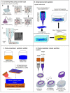
(A) Printing of GELMA-alginate blend using coaxial nozzle with simultaneous crosslinking. Reproduced with permission form [34]. Copyright 2016, John Wiley and Sons; (B) Temperature sensitive gelation of gelatin alginate bioink; (C) Photo-crosslinked PCL scaffold with gelatin methacrylamide; (D) Photo-crosslinked natural HAMA scaffold for ring structure.
Figure 4
Figure 4
(A) Design of a fiber reinforced single tube (inner diameter 4 mm, outer diameter 6 mm, length 20 mm) including anchoring stands (white: PCL, red: Gelma-gellan, blue: alginate); (B) construct directly after printing, scalpel cuts for removing stands represented by dashed lines; (C) tube after removing PCL stands; (D) cross-section of printed tube, right: design, left: after infusion with gelatin containing MSCs (safranin-O staining, red: Gelma-gellan tube wall; green: Gelatin-MSC mixture). Reinforcing PCL fibers that were present in the tube wall were dissolved during the embedding process; (E) magnification from picture D of gelatin hydrogel containing MSCs (blue dots) (scale bar represents 200 µm). Reproduced with permission from [65]. Copyright 2016, IOP Publishing; (B) Micro-carrier printing in GELMA bioink. Reproduced with permission from [35]. Copyright 2016, IOP Publishing.; (C) HepG2 spheroid printing using GELMA bioink for perfusion bioreactor. Reproduced with permission from [36]. Copyright 2016, IOP Publishing.
Figure 5
Figure 5
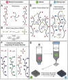
(A) Polymer or polymer mixtures can be linear (e.g., gelatin), branched (e.g., 4 arm PEG amine), or multifunctional (e.g., gelatin methacrylate); (B) PEGX can be linear or multiarm and can be various chain lengths; (C) Cells can be optionally incorporated by (D) mixing with polymers and PEGX to form the bioink; (E) Optional, secondary crosslinking to increase mechanical robustness may be performed postprinting; (F) By changing the reactive groups of PEGX, polymers of other functional groups may be crosslinked; (G) Printing process of PEGX bioink method and corresponding phase: PEGX with or without cells are mixed within the polymer solution and loaded into the printing cartridge. After gel formation and stable mechanical properties are achieved, gels can be 3D printed into multilayer structures. Pink: Bioink formulation, Green: Bioink, Blue: Robust gel formed after crosslinking. Reproduced with permission from [33]. Copyright 2016, John Wiley and Sons.
Figure 6
Figure 6
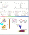
(A) (a) Work flow of nanostructured pluronic bioink: Pluronic is mixed with cells and other polymers such as hyaluronic acid methacrylate at a low temperature (handling regime) and is then formed to a physical gel for the printing process via temperature gelation. Subsequent UV crosslinking is then performed for mechanical stability. The crosslinked gel is washed to introduce the nanostructures and reduce the total pluronic concentration for the cell culture. (b) Chemical structure of pluronic F127 before and after the reaction with acryloyl chloride. Reproduced with permission from [55]. Copyright 2016, IOP Publishing; (B) Schematic illustration of a M13 recombinant DNA, RGD and calcium binding domains and bioink (target cells + RGD-phages + alginate) with a 3-D cell-laden scaffold printed using the phage-based bioink.
Figure 7
Figure 7
Effect of shear stress on Newtonian and non-Newtonian bioinks and distribution of shear stress and pressure distribution inside syringe.
Figure 8
Figure 8
(A) Hyaluronic acid modified with both methacrylates (blue) and guest-host molecules (purple). Ad–MeHA and CD–MeHA macromers crosslink by both physical bonding upon mixing and through a secondary crosslinking of methacrylates with UV light exposure. (B) The printing of channels, by writing an ink into a support gel that is modified for secondary crosslinking. UV irradiation covalently crosslinks the support gel around the ink. Pressure driven flow results in removal of the ink, leaving a channel network (C) The printing of self-supporting structures, by writing an ink that can be covalently crosslinked into a support gel, followed by UV crosslinking, and dissolution of the support with excess β-CD. Scalebars: 500 μm. Reproduced with permission from [22]. Copyright 2016, John Wiley and Sons.

References

    1. Mironov V., Boland T., Trusk T., Forgacs G., Markwald R.R. Organ printing: Computer-aided jet-based 3D tissue engineering. Trends Biotechnol. 2003;21:157–161. doi: 10.1016/S0167-7799(03)00033-7. - DOI - PubMed
    1. Chang R., Nam J., Sun W. Direct cell writing of 3D microorgan for in vitro pharmacokinetic model. Tissue Eng. Part C Methods. 2008;14:157–166. doi: 10.1089/ten.tec.2007.0392. - DOI - PubMed
    1. Huh D., Hamilton G.A., Ingber D.E. From 3D cell culture to organs-on-chips. Trends Cell Biol. 2011;21:745–754. doi: 10.1016/j.tcb.2011.09.005. - DOI - PMC - PubMed
    1. Derby B. Printing and prototyping of tissues and scaffolds. Science. 2012;338:921–926. doi: 10.1126/science.1226340. - DOI - PubMed
    1. Zhang Z., Cui H. Biodegradability and biocompatibility study of poly(chitosan-g-lactic acid) scaffolds. Molecules. 2012;17:3243–3258. doi: 10.3390/molecules17033243. - DOI - PMC - PubMed