. 2016 May 24;8(6):318.
doi: 10.3390/nu8060318.

Cuminaldehyde from Cinnamomum verum Induces Cell Death through Targeting Topoisomerase 1 and 2 in Human Colorectal Adenocarcinoma COLO 205 Cells

Affiliations

Cuminaldehyde from Cinnamomum verum Induces Cell Death through Targeting Topoisomerase 1 and 2 in Human Colorectal Adenocarcinoma COLO 205 Cells

Kuen-Daw Tsai et al. Nutrients. .

Abstract

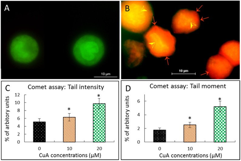
Cinnamomum verum, also called true cinnamon tree, is employed to make the seasoning cinnamon. Furthermore, the plant has been used as a traditional Chinese herbal medication. We explored the anticancer effect of cuminaldehyde, an ingredient of the cortex of the plant, as well as the molecular biomarkers associated with carcinogenesis in human colorectal adenocarcinoma COLO 205 cells. The results show that cuminaldehyde suppressed growth and induced apoptosis, as proved by depletion of the mitochondrial membrane potential, activation of both caspase-3 and -9, and morphological features of apoptosis. Moreover, cuminaldehyde also led to lysosomal vacuolation with an upregulated volume of acidic compartment and cytotoxicity, together with inhibitions of both topoisomerase I and II activities. Additional study shows that the anticancer activity of cuminaldehyde was observed in the model of nude mice. Our results suggest that the anticancer activity of cuminaldehyde in vitro involved the suppression of cell proliferative markers, topoisomerase I as well as II, together with increase of pro-apoptotic molecules, associated with upregulated lysosomal vacuolation. On the other hand, in vivo, cuminaldehyde diminished the tumor burden that would have a significant clinical impact. Furthermore, similar effects were observed in other tested cell lines. In short, our data suggest that cuminaldehyde could be a drug for chemopreventive or anticancer therapy.

Keywords: antiproliferative; cuminaldehyde; lysosomal vacuolation; topoisomerase I; topoisomerase II; xenograft.


Figures

Figure 1
Figure 1
Cuminaldehyde’s chemical structure and effects on cellular morphology, proliferation, as well as lactate dehydrogenase releasing in human colorectal COLO 205 cells. (A) Chemical structure; (B) and (C) Cuminaldehyde’s effect on cellular morphology; Cells were treated without (B) and with 20 μM (C) cuminaldehyde for 48 h. Cell detachment, shrinkage, and blebbing of plasma membrane (arrows) were found when the cells were incubated with 20 μM of cuminaldehyde; (D) Cuminaldehyde’s effect on growth. Human colorectal COLO 205 cells were treated with cuminaldehyde at the specified circumstances. Proliferation suppressive effect was determined using the XTT test; (E) Cuminaldehyde’s effect on the lactate dehydrogenase releasing in the cells. The supernatant was gathered after 48 h of incubation with the indicated cuminaldehyde concentrations. Absorptions of light were determined by a spectrophotometer (Tecan infinite M200, Tecan, Männedorf, Switzerland). Results are shown by means plus/minus standard error of the mean, n equal to 3. *, Statistically significant (p less than 0.05) from the control group. CuA, cuminaldehyde.
Figure 2
Figure 2
Cuminaldehyde caused nuclear fragmentation in human colorectal COLO 205 cells. (A and B) Acridine orange staining; COLO 205 cells were incubated without (A) with 20 μM (B) cuminaldehyde, respectively, for 48 h, then stained using acridine orange. The orange vacuoles in COLO cells demonstrate that they existed acidic; (A) Typical picture of control cells accompanying intact nucleus with green fluorescence that implicates a good cell viability; (B) Typical picture of test cells incubated with cuminaldehyde with lysosomal vacuolation (arrows) and nuclear fragmentation (arrow heads) were found; (C and D) Comet test. Cuminaldehyde’s effect on intensities of tail (C) as well as moment (D). Human colorectal COLO 205 cells were incubated with cuminaldehyde at the indicated concentrations for 48 h. Data are shown as means plus/minus standard error of the mean, n = 125. *, Significant difference (p < 0.05) from the control. CuA, cuminaldehyde.
Figure 3
Figure 3
Cuminaldehyde increased the volume of the acidic compartment in human colorectal COLO 205 cells. After treatment without and with 20 μM cuminaldehyde, respectively, for 48 h, human colorectal COLO 205 cells were stained using neutral red. (A) Human colorectal COLO 205 cells without treatment: There were no observable vacuoles in the cell; (B) Human colorectal COLO 205 cells treated with cuminaldehyde at the concentration of 20 μM for 48 h. The blebbing (black arrows) and acidic red-stained vacuoles (red arrows) in cells happened; (C) Cuminaldehyde increased volume of acidic compartment in a quantity-dependent manner. After treating the cells using the specified concentrations of cuminaldehyde for 48 h, results were evaluated by a spectrophotometer. Results are shown by means plus/minus standard error of the mean, n equal to 3. *, Statistically significant (p less than 0.05) from the control group. CuA, cuminaldehyde.
Figure 4
Figure 4
Cuminaldehyde caused apoptosis via the mitochondrial pathway in human colorectal COLO 205 cells. (A) Cells were treated with the specified cuminaldehyde concentrations for 48 h and mitochondrial membrane potential was evaluated using JC-1 spectrophotometrically; (B) Activations of caspase-3 as well as -9. After treating the cells using the specified concentrations of cuminaldehyde for 48 h, activities of caspases-3 and -9 were determined using a spectrophotometer. Results are expressed by means plus/minus standard error of the mean, n equal to 3. *, Statistically significant (p less than 0.05) from the control group. CuA, cuminaldehyde.
Figure 5
Figure 5
Cuminaldehyde inhibited topoisomerase I as well as II activities in human colorectal COLO 205 cells. (A) Cuminaldehyde inhibited topoisomerase I activity. Nuclear proteins of COLO 205 cells interacted with the indicated cuminaldehyde concentrations in a topoisomerase I’s specific reaction mixture (lanes 3–5), or 60 μM of camptothecin (CPT, a specific topoisomerase I inhibitor and used as positive control, lane 6), or the vehicle (1% dimethyl sulfoxide, lane 2). Lane 1, pUC19 DNA only; (B) Cuminaldehyde inhibited topoisomerase II activity. DNA relaxation test (upper panel) and decatenation test (lower panel). Nuclear proteins of COLO 205 cells were added to a specific topoisomerase II reaction mixture with the specified cuminaldehyde concentrations (lanes 3–5) or 60 μM of camptothecin (a specific suppressor of topoisomerase II and used as positive control, lane 6), or the vehicle (one percent dimethyl sulfoxide, lane 2). Lane 1, Interwined pUC19 DNA (upper panel) or kinetoplast DNA (lower panel) only. kinetoplast DNA is an extensive chain of plasmids. When kinetoplast DNA is examined using electrophoretic analysis, it gets the gel only a lightly (figure not demonstrated). Consequent to topoisomerase II’s decatenation, small monomeric circles of nucleic acid were produced (lower panel, lane 2–6). This is the representative of six experiments. CPT, camptothecin; CuA, cuminaldehyde; kDNA, kinetoplast; S & R, Interwined and the unrestrained forms of the pUC 19 plasmid, respectively; VP-16, etoposide.
Figure 6
Figure 6
Cuminaldehyde suppressed growth and caused apoptosis in human colorectal COLO 205 xenograft. The mice with pre-established cancers (n = 8 per group) were treated using intratumoral injection with the specified cuminaldehyde concentrations. Tumor volumes were recorded by calipers and apoptosis was evaluated by terminal deoxynucleotidyl transferase dUTP nick end labeling test. (A) Left panel, Representative of tumor-bearing mice from the control (orange arrows) and 20 mg/kg/day of cuminaldehyde-injected (blue arrows) groups; (A) Right panel, cuminaldehyde caused apoptosis in human colorectal COLO 205 xenograft using terminal deoxynucleotidyl transferase dUTP nick end labeling test. Representative of terminal deoxynucleotidyl transferase dUTP nick end labeling test of tumors from the control (white arrows) and 20 mg/kg/day of cuminaldehyde-injected (yellow arrows) groups; (B) Mean of tumor volume observed at the specified number of days after the start of treatment; (C) Cuminaldehyde’s effects on tumor weight observed at the endpoint of the experiment. Tumor weight per mouse was collected and analyzed. Results are shown by means plus/minus standard error of the mean, n = 8. *, Statistically significant (p less than 0.05) from the control group. CuA, cuminaldehyde.

References

    1. Tanzer M., Liebl M., Quante M. Molecular biomarkers in esophageal, gastric, and colorectal adenocarcinoma. Pharmacol. Ther. 2013;140:133–147. doi: 10.1016/j.pharmthera.2013.06.005. - DOI - PubMed
    1. Tanaka S., Yoon Y.H., Fukui H., Tabata M., Akira T., Okano K., Iwai M., Iga Y., Yokoyama K. Antiulcerogenic compounds isolated from chinese cinnamon. Planta Medica. 1989;55:245–248. doi: 10.1055/s-2006-961994. - DOI - PubMed
    1. Reddy A.M., Seo J.H., Ryu S.Y., Kim Y.S., Kim Y.S., Min K.R., Kim Y. Cinnamaldehyde and 2-methoxycinnamaldehyde as NF-κB inhibitors from Cinnamomum cassia. Planta Medica. 2004;70:823–827. doi: 10.1055/s-2004-827230. - DOI - PubMed
    1. Shukla S., Meeran S.M., Katiyar S.K. Epigenetic regulation by selected dietary phytochemicals in cancer chemoprevention. Cancer Lett. 2014;355:9–17. doi: 10.1016/j.canlet.2014.09.017. - DOI - PMC - PubMed
    1. Priyadarsini R.V., Nagini S. Cancer chemoprevention by dietary phytochemicals: Promises and pitfalls. Curr. Pharm. Biotechnol. 2012;13:125–136. doi: 10.2174/138920112798868610. - DOI - PubMed

MeSH terms