. 2016 May 27;12(5):e1006100.
doi: 10.1371/journal.pgen.1006100. eCollection 2016 May.

The Gene Expression Program for the Formation of Wing Cuticle in Drosophila

Affiliations

The Gene Expression Program for the Formation of Wing Cuticle in Drosophila

Lukasz F Sobala et al. PLoS Genet. .

Abstract

The cuticular exoskeleton of insects and other arthropods is a remarkably versatile material with a complex multilayer structure. We made use of the ability to isolate cuticle synthesizing cells in relatively pure form by dissecting pupal wings and we used RNAseq to identify genes expressed during the formation of the adult wing cuticle. We observed dramatic changes in gene expression during cuticle deposition, and combined with transmission electron microscopy, we were able to identify candidate genes for the deposition of the different cuticular layers. Among genes of interest that dramatically change their expression during the cuticle deposition program are ones that encode cuticle proteins, ZP domain proteins, cuticle modifying proteins and transcription factors, as well as genes of unknown function. A striking finding is that mutations in a number of genes that are expressed almost exclusively during the deposition of the envelope (the thin outermost layer that is deposited first) result in gross defects in the procuticle (the thick chitinous layer that is deposited last). An attractive hypothesis to explain this is that the deposition of the different cuticle layers is not independent with the envelope instructing the formation of later layers. Alternatively, some of the genes expressed during the deposition of the envelope could form a platform that is essential for the deposition of all cuticle layers.


Conflict of interest statement

The authors have declared that no competing interests exist.

Figures

Fig 1
Fig 1. Changes in pupal wing morphology over time in wild type and mutants.
Shown are wing discs and pupal wings from wild type (Ore-R (A-E), dyl-kd (F-J), ect-kd (K-O) and CG8213-kd (P-T) animals. The wing discs were dissected from wandering third instar larvae and during this period the disc prepares for evagination. This leads to some discs being a bit more 3 dimensional (this can be seen in panel F). The folding pattern seen in post-flattening/expansion wild type pupal wing discs is highly reproducible. The mutants are more variable from animal to animal. This was particularly true for ect, where males showed a stronger phenotype than females. The final column shows pigmented pupal wings imaged through the pupal case and establishes that the differences between wild type and mutants are not due to dissection. The arrowheads point to shallower than normal proximal anterior folds and the arrows to ectopic folds seen in mutants. Note that during the process of dissection there is often some relaxation in the folding pattern of younger (pre-pigmentation) pupal wings.
Fig 2
Fig 2. Transmission electron micrographs of wing cuticle deposition.
Panels A-F are all shown at the same magnification. The time is for pupae collected as white prepupae and aged at 25°C. Panel A. In 42 hr awp wings the deposition of the envelope (en) is visible (arrows). Gaps remain where the envelope does not cover the surface (asterisk). B. In 52 hr awp wings the epicuticle (epi) is now visible (line) and pore canals are prominent (arrows—pc). C. In 62 hr wings procuticle is now obvious (line). Pore canals remain prominent (arrow) and at the base of the procuticle a dark granular layer is first seen (asterisk). This may be the adhesion layer. D. In 72 hr wings the procuticle (pro) is prominent (line) and the dark granular layer is more obvious (asterisk). Panel E. In the 88 hr wing the procuticle (line) and dark granular layer (asterisk) are both prominent. Panel F. In the 96 hr wing the procuticle (line) and dark granular layer (asterisk) are both prominent. Panels G-J are relatively low magnification electron micrographs of 76–80 hr pupal wings where both the dorsal and ventral cuticle can be seen. In all of these images the putative dorsal surface is marked by an arrow. G. Ore-R. H. ap>dyl RNAi. I. ap>ect RNAi. J. ap>CG8213 RNAi. Note in all three of these images there are gaps between the apical surface of the dorsal wing cells and the cuticle (asterisks) and the dorsal and ventral cuticle is of different thickness in H-J compared to G. In panel G the arrow also points to a hair pedestal. Panels K and L show regions of highly abnormal cuticle in ap>dyl RNAi wings. The arrows point to gaps/holes in the procuticle. M shows a region of an ap>CG8213 RNAi wing with abnormal procuticle (arrows).
Fig 3
Fig 3. Gene expression clusters during the deposition of wing cuticle.
A. The dendrogram shows the relationships between the gene expression patterns at different time points. The scale refers directly to the mutual Jensen-Shannon distance between branches. B. The gene expression patterns for all of the 16 clusters. The numbers on the Y axis show the log10(FPKM+1) values and the X axis shows the time. The graphs show the expression patterns for the members of each of the 16 clusters. Clustering methods are described in more detail in S1 File (Supplementary Methods). Colored lines represent the pattern for each gene and the black line represents the medoid.
Fig 4
Fig 4. Similar results are obtained by both RNAseq and RT-qPCR.
A. The relative gene expression patterns for the two mRNAs encoded by the CG1005 gene. Note the similar pattern seen with both RNAseq and RT-qPCR. B. The expression pattern for dyl and two isoforms of Cht6. Note that Cht6-RG expression follows a similar pattern to dyl. C. The expression patterns for two isoforms of mwh and CG14257 show qualitatively similar changes when assayed by both RNAseq and RT-qPCR. Note that relative expression values as a function of time and not absolute expression values are shown in this figure.
Fig 5
Fig 5. Heat maps for 4 groups of related genes.
A. Annotated cuticle proteins. B. Genes with a known function in cuticle deposition or maturation. C. ZP domain protein encoding genes. D. Genes that regulate transcription.
Fig 6
Fig 6. Isoform switching between neighboring time points.
A. A heat map is shown for selected genes where we detected putative switching between isoforms. Several of these genes are known/likely to have a role in cuticle formation and or wing development. Among the most interesting were the isoform shifts for Cht6 (which encodes a chitinase), Cda5 (which encodes a chitin deacetylase), laccase (which encodes an oxidase with a role in cuticle formation [40, 57, 58]) and mwh (which regulates wing hair morphogenesis [55, 56]). B. The time course for the expression of the pale isoforms is shown. Note how the 96 hr peak of expression is restricted to the epidermal form.
Fig 7
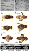
Fig 7. Phenotypes associated with 42 hr genes.
A and B show the dorsal notum of Ore-R and ap>CG8213. apterous-Gal4 drives expression in the cells that form the dorsal surface of the wing but not those that form the ventral surface. It also drives expression in the dorsal thorax (notum). Panels C-H show adult flies where a 42 hr gene was knocked down using ap-Gal4. Ore-R is shown (C) for comparison. I and J show a thoracic macrochaete from Ore-R and from ap>CG8213. Panels K-N show unmounted wings from Ore-R or knocked down 42 hr genes. Note the curved kd wings.
Fig 8
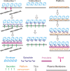
Fig 8. Models to explain the procuticle phenotypes of 42 hr genes.
In the Instructive 1 model the 42 hr proteins form the envelope, which signals back to the cell to organize the cytoskeleton so that the epicuticle and procuticle is secreted in the proper pattern. In Instructive 2 the 42 hr proteins form the envelope and when the proteins that form the epicuticle are secreted they bind to and are patterned by envelope and 42 hr proteins. A similar situation could result in the epicuticle patterning the procuticle. In the Platform model the 42 hr proteins are not part of the envelope but they form a complex that is essential for the patterned secretion of the proteins that form the 3 cuticular layers. Our second model is that many of the 42 hr genes do not encode proteins that are part of the envelope. Rather they would form a “platform” or complex that in some way mediates the tight juxtaposition of the cuticle and the apical surface of the epithelial cells and that this platform is needed for the proper deposition of the envelope and other cuticular components [22].

Similar articles

Cited by

References

    1. Vincent JF, Wegst UG. Design and mechanical properties of insect cuticle. Arthropod structure & development. 2004;33(3):187–99. - PubMed
    1. Moussian B, Veerkamp J, Muller U, Schwarz H. Assembly of the Drosophila larval exoskeleton requires controlled secretion and shaping of the apical plasma membrane. Matrix biology: journal of the International Society for Matrix Biology. 2007;26(5):337–47. - PubMed
    1. Payre F. Genetic control of epidermis differentiation in Drosophila. The International journal of developmental biology. 2004;48(2–3):207–15. - PubMed
    1. Vincent JF. Deconstructing the design of a biological material. Journal of theoretical biology. 2005;236(1):73–8. - PubMed
    1. Grunenfelder LK, Herrera S, Kisailus D. Crustacean-derived biomimetic components and nanostructured composites. Small. 2014;10(16):3207–32. 10.1002/smll.201400559 - DOI - PubMed

Publication types

MeSH terms

LinkOut - more resources