. 2016 May 27;11(5):e0156293.
doi: 10.1371/journal.pone.0156293. eCollection 2016.

Cryopreservation of Human Mucosal Leukocytes

Affiliations

Cryopreservation of Human Mucosal Leukocytes

Sean M Hughes et al. PLoS One. .

Abstract

Background: Understanding how leukocytes in the cervicovaginal and colorectal mucosae respond to pathogens, and how medical interventions affect these responses, is important for developing better tools to prevent HIV and other sexually transmitted infections. An effective cryopreservation protocol for these cells following their isolation will make studying them more feasible.

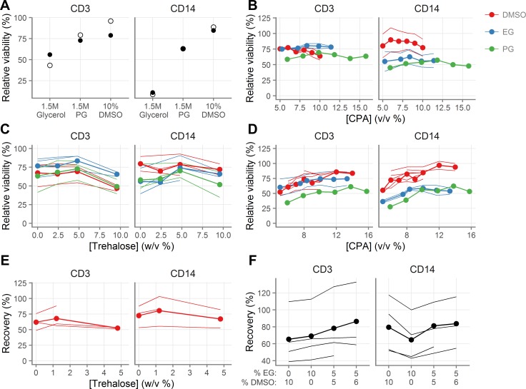
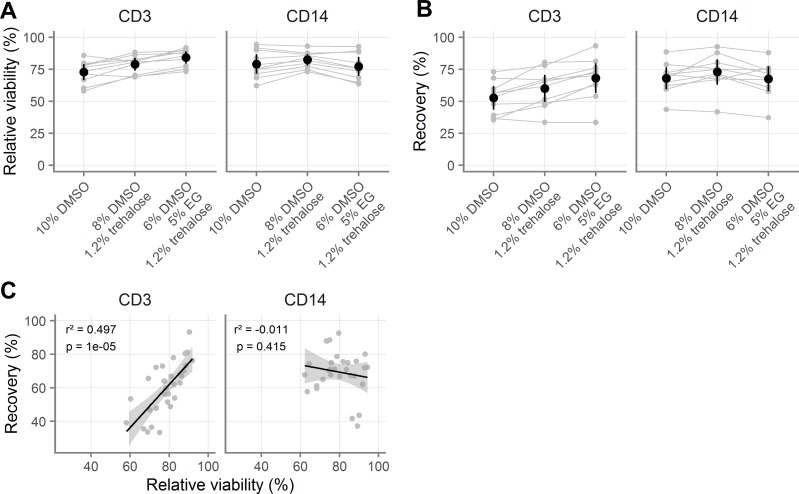
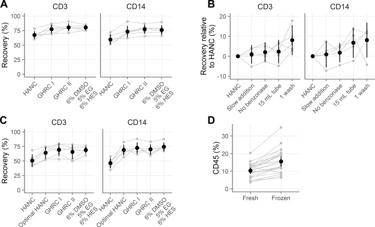
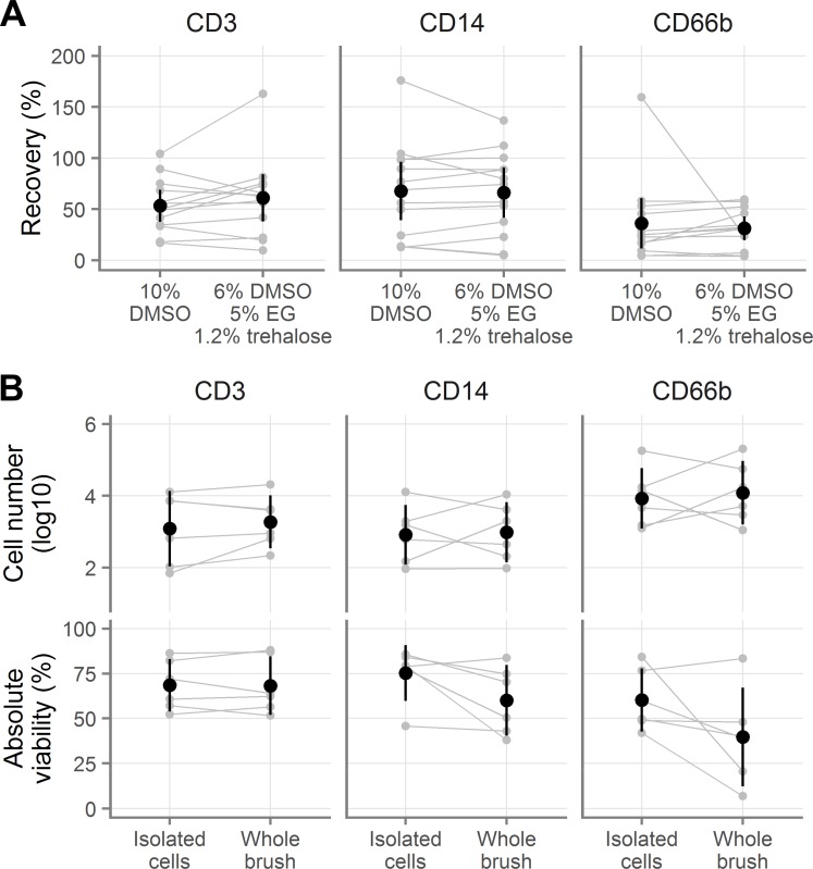
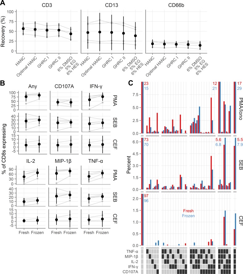
Methods and findings: To find an optimal cryopreservation protocol for mucosal mononuclear leukocytes, we compared cryopreservation media and procedures using human vaginal leukocytes and confirmed our results with endocervical and colorectal leukocytes. Specifically, we measured the recovery of viable vaginal T cells and macrophages after cryopreservation with different cryopreservation media and handling procedures. We found several cryopreservation media that led to recoveries above 75%. Limiting the number and volume of washes increased the fraction of cells recovered by 10-15%, possibly due to the small cell numbers in mucosal samples. We confirmed that our cryopreservation protocol also works well for both endocervical and colorectal leukocytes. Cryopreserved leukocytes had slightly increased cytokine responses to antigenic stimulation relative to the same cells tested fresh. Additionally, we tested whether it is better to cryopreserve endocervical cells on the cytobrush or in suspension.

Conclusions: Leukocytes from cervicovaginal and colorectal tissues can be cryopreserved with good recovery of functional, viable cells using several different cryopreservation media. The number and volume of washes has an experimentally meaningful effect on the percentage of cells recovered. We provide a detailed, step-by-step protocol with best practices for cryopreservation of mucosal leukocytes.


Conflict of interest statement

Competing Interests: The authors have declared that no competing interests exist.

Figures

Fig 1
Fig 1. Development of novel cryopreservation medium.
A, Viability of vaginal T cells (indicated “CD3”) and macrophages (indicated “CD14”) following cryopreservation with 1.5M glycerol, 1.5M propylene glycol (PG) or 10% dimethyl sulfoxide (DMSO). B, Effect of CPA concentration on cell viability after cryopreservation. C, Effect of trehalose concentration on cell viability after cryopreservation in the presence 0.84M DMSO, 1.5M PG, or 1.75M EG. D, Effect of CPA concentration on cell viability after cryopreservation in the presence of 4.8% trehalose. E, Effect of trehalose concentration on recovery after cryopreservation in the presence of 1.4M DMSO. F, Effect of mixtures of DMSO and EG on cell recovery after cryopreservation in the presence of 1.2% trehalose. Each line for a given color indicates a different tissue sample. The line with the circles indicates the mean across the different tissue samples. Measurements were made in duplicate for each tissue sample. For relative viability, 100% indicates the same viability as the fresh cells.
Fig 2
Fig 2. Validation of novel cryopreservation medium.
A-B, Comparison of three cryopreservation media by relative viability (A) and recovery (B) for vaginal T cells (indicated “CD3”) and macrophages (indicated “CD14”). All samples were processed with procedure A (see Table 1). C, Correlation of relative viability and recovery for vaginal T cells and macrophages with different cryomedia. For relative viability, 100% indicates the same viability as the fresh cells. Gray symbols indicate the average of duplicates from the same sample, with gray lines indicating pairing. Black symbols show the mean across all samples and black vertical lines show the 95% confidence interval of the mean. In C, black line indicates the slope of the linear model and the grey shaded area indicates the 95% confidence interval. Adjusted p-values and r2 values are shown from linear regression.
Fig 3
Fig 3. Comparison of novel cryopreservation medium with published methods.
A, Recovery of vaginal T cells (indicated “CD3”) and macrophages (indicated “CD14”) with several cryopreservation media. GHRC I [Global HIV Vaccine Research Cryorepository] is 10% DMSO alone and GHRC II is 5% DMSO with 6% hydroxyethyl starch, both in RPMI with 12.5% bovine serum albumin. HANC is 10% DMSO in FBS. HANC was processed according to procedure C and the others were processed with procedure B (see Table 1). B, Evaluation of which processing steps improve the recovery obtained relative to the standard HANC procedure, where positive values indicate improvements over HANC. C, Rescue of HANC procedure (“optimal HANC”) by use of optimal processing procedures for small sample sizes. D, Percentage of all vaginal cells that are CD45+ before and after freezing. Gray symbols indicate the average of duplicates, with gray lines indicating pairing. Black symbols show the mean across all samples and black vertical lines show the 95% confidence interval of the mean.
Fig 4
Fig 4. Cryopreservation of cells derived from endocervical cytobrushes.
A, Recovery of endocervical T cells (indicated “CD3”), macrophages (indicated “CD14”), and neutrophils (indicated “CD66b”), when cryopreserved after isolation from cytobrushes. B, Absolute number of live (top) and viability of total endocervical cells (bottom) after cryopreservation either as a cell suspension or still on the cytobrush. Gray symbols indicate the individual samples, with gray lines indicating pairing. Black symbols show the mean across all samples and black vertical lines show the 95% confidence interval of the mean.
Fig 5
Fig 5. Recovery and functionality of colorectal cells after cryopreservation.
A, Recovery of colorectal T cells (indicated “CD3”), macrophages (indicated “CD13”), and neutrophils (indicated “CD66b”), after cryopreservation with several cryopreservation media. GHRC I [Global HIV Vaccine Research Cryorepository] is 10% DMSO alone and GHRC II is 5% DMSO with 6% hydroxyethyl starch, both in RPMI with 12.5% bovine serum albumin. HANC is 10% DMSO in FBS. HANC was processed according to procedure C and the others were processed with procedure B (see Table 1). B, Background-subtracted cytokine production from colorectal CD8 T cells fresh or after cryopreservation after stimulation with phorbol 12-myristate 13-acetate (PMA); staphylococcal enterotoxin B (SEB); or human cytomegalovirus, Epstein-Barr virus and influenza virus peptides (CEF). Gray symbols indicate the individual samples, with gray lines indicating pairing. Black symbols show the mean across all samples and black vertical lines show the 95% confidence interval of the mean. C, Polyfunctionality of colorectal CD8 cytokine responses to stimulation. Red bars indicate fresh samples and blue bars indicate samples tested after cryopreservation. Each bar corresponds to the percent of CD8 cells that expressed the indicated cytokines, averaged across the donors. Percentages are indicated explicitly for bars that exceed the axis limits of the graphs. In the legend, a dark square indicates presence of that cytokine and a light square its absence. White gaps divide the graphs and legends into groups expressing 0, 1, 2, 3, 4, or 5 cytokines.

References

    1. Liebenberg LJ, Gamieldien H, Mkhize NN, Jaumdally SZ, Gumbi PP, Denny L, et al. Stability and transport of cervical cytobrushes for isolation of mononuclear cells from the female genital tract. J Immunol Methods. Elsevier B.V.; 2011;367: 47–55. 10.1016/j.jim.2011.01.013 - DOI - PMC - PubMed
    1. Bull M, Lee D, Stucky J, Chiu Y-L, Rubin A, Horton H, et al. Defining blood processing parameters for optimal detection of cryopreserved antigen-specific responses for HIV vaccine trials. J Immunol Methods. 2007;322: 57–69. 10.1016/j.jim.2007.02.003 - DOI - PMC - PubMed
    1. Mazur P. Kinetics of Water Loss From Cells At Subzero Temperatures and the Likelihood of Intracellular Freezing. J Gen Physiol. 1963;47: 347–369. 10.1085/jgp.47.2.347 - DOI - PMC - PubMed
    1. Shu Z, Hughes SM, Fang C, Huang J, Fu B, Zhao G, et al. A Study of the Osmotic Characteristics, Water Permeability, and Cryoprotectant Permeability of Human Vaginal Immune Cells. Cryobiology. 2016;72:93–9. 10.1016/j.cryobiol.2016.03.003 - DOI - PMC - PubMed
    1. Shu Z, Hughes SM, Fang C, Hou Z, Zhao G, Fialkow M, et al. Determination of the Membrane Permeability to Water of Human Vaginal Mucosal Immune Cells at Subzero Temperatures Using Differential Scanning Calorimetry. Biopreserv Biobank. 2016; bio.2015.0079. 10.1089/bio.2015.0079 - DOI - PMC - PubMed