Protein Disulfide Levels and Lens Elasticity Modulation: Applications for Presbyopia
- PMID: 27233034
- PMCID: PMC5995025
- DOI: 10.1167/iovs.15-18413
Protein Disulfide Levels and Lens Elasticity Modulation: Applications for Presbyopia
Abstract
Purpose: The purpose of the experiments described here was to determine the effects of lipoic acid (LA)-dependent disulfide reduction on mouse lens elasticity, to synthesize the choline ester of LA (LACE), and to characterize the effects of topical ocular doses of LACE on mouse lens elasticity.
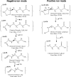
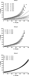
Methods: Eight-month-old mouse lenses (C57BL/6J) were incubated for 12 hours in medium supplemented with selected levels (0-500 μM) of LA. Lens elasticity was measured using the coverslip method. After the elasticity measurements, P-SH and PSSP levels were determined in homogenates by differential alkylation before and after alkylation. Choline ester of LA was synthesized and characterized by mass spectrometry and HPLC. Eight-month-old C57BL/6J mice were treated with 2.5 μL of a formulation of 5% LACE three times per day at 8-hour intervals in the right eye (OD) for 5 weeks. After the final treatment, lenses were removed and placed in a cuvette containing buffer. Elasticity was determined with a computer-controlled instrument that provided Z-stage upward movements in 1-μm increments with concomitant force measurements with a Harvard Apparatus F10 isometric force transducer. The elasticity of lenses from 8-week-old C57BL/6J mice was determined for comparison.
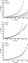
Results: Lipoic acid treatment led to a concentration-dependent decrease in lens protein disulfides concurrent with an increase in lens elasticity. The structure and purity of newly synthesized LACE was confirmed. Aqueous humor concentrations of LA were higher in eyes of mice following topical ocular treatment with LACE than in mice following topical ocular treatment with LA. The lenses of the treated eyes of the old mice were more elastic than the lenses of untreated eyes (i.e., the relative force required for similar Z displacements was higher in the lenses of untreated eyes). In most instances, the lenses of the treated eyes were even more elastic than the lenses of the 8-week-old mice.
Conclusions: As the elasticity of the human lens decreases with age, humans lose the ability to accommodate. The results, briefly described in this abstract, suggest a topical ocular treatment to increase lens elasticity through reduction of disulfides to restore accommodative amplitude.
Figures


References
-
- Weeber HA, van der Heijde RGL. . Internal deformation of the human crystalline lens during accommodation. Acta Ophthalmol. 2008; 86: 642– 647. - PubMed
-
- Farnsworth PN, Mauriello JA, Burke-Gadomski P, Kulyk T, Cinotti AA. . Surface ultrastructure of the human lens capsule and zonular attachments. Invest Ophthalmol. 1976; 15: 36– 40. - PubMed
-
- Hiraoka M, Inoue K-I, Ohtaka-Maruyama C,et al. . Intracapsular organization of ciliary zonules in monkey eyes. Anat Rec. 2010; 293: 1797– 1804. - PubMed
Publication types
MeSH terms
Substances
Grants and funding
LinkOut - more resources
Full Text Sources
Other Literature Sources
Research Materials
Miscellaneous

