Review
. 2016 Dec;73(24):4577-4590.
doi: 10.1007/s00018-016-2282-2. Epub 2016 May 28.

Mitochondrial cAMP signaling

Affiliations
Review

Mitochondrial cAMP signaling

Fan Zhang et al. Cell Mol Life Sci. 2016 Dec.

Abstract

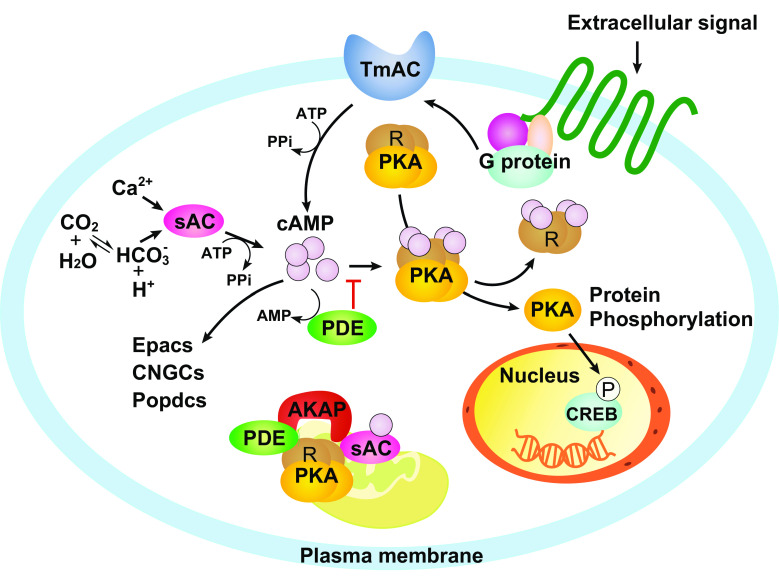
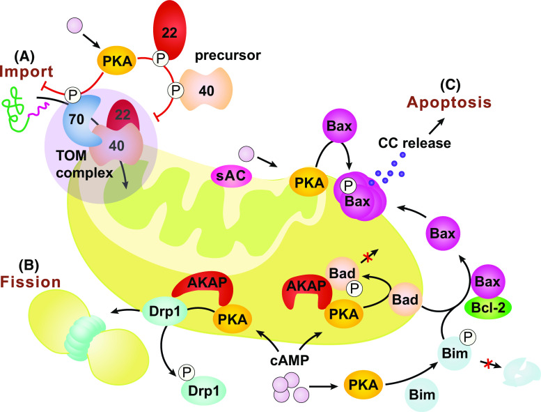
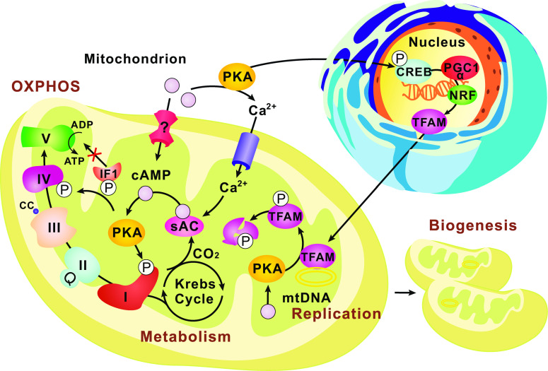
Cyclic adenosine 3, 5'-monophosphate (cAMP) is a ubiquitous second messenger regulating many biological processes, such as cell migration, differentiation, proliferation and apoptosis. cAMP signaling functions not only on the plasma membrane, but also in the nucleus and in organelles such as mitochondria. Mitochondrial cAMP signaling is an indispensable part of the cytoplasm-mitochondrion crosstalk that maintains mitochondrial homeostasis, regulates mitochondrial dynamics, and modulates cellular stress responses and other signaling pathways. Recently, the compartmentalization of mitochondrial cAMP signaling has attracted great attentions. This new input should be carefully taken into account when we interpret the findings of mitochondrial cAMP signaling. In this review, we summarize previous and recent progress in our understanding of mitochondrial cAMP signaling, including the components of the signaling cascade, and the function and regulation of this signaling pathway in different mitochondrial compartments.

Keywords: AKAP; BH3; Drp1; Fission; PKA; Protein import; TFAM; mtDNA.


Figures

Fig. 1
Fig. 1
General cAMP signaling pathways. Intracellular cAMPs are generated by two classes of ACs, the transmembrane AC (TmAC) and the soluble AC (sAC). Both TmAC and sAC convert ATP to cAMP upon stimulus. TmAC can be activated by G proteins when an extracellular signal is received by the G protein-coupled membrane receptor. sAC is insensitive to G-proteins but can be activated by bicarbonate and calcium. Elevated cAMP level activates protein kinase A (PKA), the major effector of cAMP signaling, by releasing the two catalytic subunits (Cs) from the two regulatory subunits (Rs). Activated PKA in turn phosphorylates and activates numerous downstream protein targets, including the cAMP response element binding protein (CREB), a transcriptional co-factor regulating multiple cellular processes. Cyclic nucleotide phosphodiesterases (PDEs) are the negative regulators that terminate cAMP-PKA signaling by hydrolyzing cAMP to AMP. As a result, cAMP level and signaling activity are determined by the equilibrium between ACs and PDEs. In addition to its main effector PKA, cAMP can also directly activate the exchange protein Epac, the cyclic nucleotide-gated channels (CNGCs) and the Popeye domain-containing proteins (Popdcs). Within a single cAMP cascade, ACs, PKA, other downstream effectors and PDEs are often tethered together by an A-kinase anchoring proteins (AKAPs) at distinct subcellular locations
Fig. 2
Fig. 2
cAMP signaling in the outer-mitochondrial compartment. cAMP from the cytosol or produced by sAC on the OMM can activate the local PKA which in turn phosphorylates different targets associated with the OMM. a PKA phosphorylation impairs the receptor activity of TOM70 and its interaction with the metabolite carrier/chaperone, prevents TOM22 translocation and TOM40 integration into the OMM, and eventually slows down the import of mitochondrial proteins. b PKA phosphorylation can also block Drp1’s translocation to the OMM surface and thus lead to reduced mitochondrial fission. c In mammals, PKA phosphorylation inhibits Bad’s apoptotic activity but promotes Bim’s by increasing its stability against proteasome-dependent degradation. PKA phosphorylation of Bax promotes its translocation to mitochondria and triggers the release of cytochrome c (CC) and the maturation of the apoptosome, which eventually leads to apoptosis. AKAPs tether PKA and other proteins, e.g., Bad, on the OMM to facilitate the cAMP-PKA targeting. They also promote different signaling specificities under the same environmental context by providing a dynamic platform of proteins complex in multiple combinations
Fig. 3
Fig. 3
cAMP signaling in the mitochondrial matrix. The intra-mitochondrial cAMP-PKA pathway has been proposed to fine-tune metabolism by directly regulating the TCA cycle and respiration. sACs have been found in the mitochondrial matrix and produce cAMP locally in response to the CO2/HCO3 generated by the TCA cycle. The matrix cAMP-PKA cascades are then activated, leading to the phosphorylation of ETC proteins such as Complex I and Complex IV subunits, modulating the OXPHOS and ATP production. In addition, PKA phosphorylation of the ATPase inhibitory factor 1 (IF1) abolishes its ability to bind to and inhibit Complex V. The sACs can also be activated by mitochondrial uptake of Ca2+. Active mechanisms for transporting cAMP into the matrix remain to be identified. To coordinate the OXPHOS and energy needs, the cAMP-PKA pathways outside mitochondria can activate the nuclear CREBs and the downstream transcription factors (PGC-1α, NRF) to promote TFAM (mtTFA) production, mtDNA replication and eventually mitochondrial biogenesis. On the other hand, the matrix cAMP signaling could exert a negative regulation on this process by increasing the PKA phosphorylation-dependent degradation of TFAM

References

    1. Wallace DC. A mitochondrial paradigm of metabolic and degenerative diseases, aging, and cancer: a dawn for evolutionary medicine. Annu Rev Genet. 2005;39:359–407. doi: 10.1146/annurev.genet.39.110304.095751. - DOI - PMC - PubMed
    1. Pallen MJ. Time to recognise that mitochondria are bacteria? Trends Microbiol. 2011;19(2):58–64. doi: 10.1016/j.tim.2010.11.001. - DOI - PubMed
    1. Vafai SB, Mootha VK. Mitochondrial disorders as windows into an ancient organelle. Nature. 2012;491(7424):374–383. doi: 10.1038/nature11707. - DOI - PubMed
    1. Chacinska A, Koehler CM, Milenkovic D, Lithgow T, Pfanner N. Importing mitochondrial proteins: machineries and mechanisms. Cell. 2009;138(4):628–644. doi: 10.1016/j.cell.2009.08.005. - DOI - PMC - PubMed
    1. Schmidt O, Pfanner N, Meisinger C. Mitochondrial protein import: from proteomics to functional mechanisms. Nat Rev Mol Cell Biol. 2010;11(9):655–667. doi: 10.1038/nrm2959. - DOI - PubMed

LinkOut - more resources