. 2017 Jan;287(Pt 4):452-460.
doi: 10.1016/j.expneurol.2016.05.031. Epub 2016 May 24.

A cognitive prosthesis for memory facilitation by closed-loop functional ensemble stimulation of hippocampal neurons in primate brain

Affiliations

A cognitive prosthesis for memory facilitation by closed-loop functional ensemble stimulation of hippocampal neurons in primate brain

Sam A Deadwyler et al. Exp Neurol. 2017 Jan.

Abstract

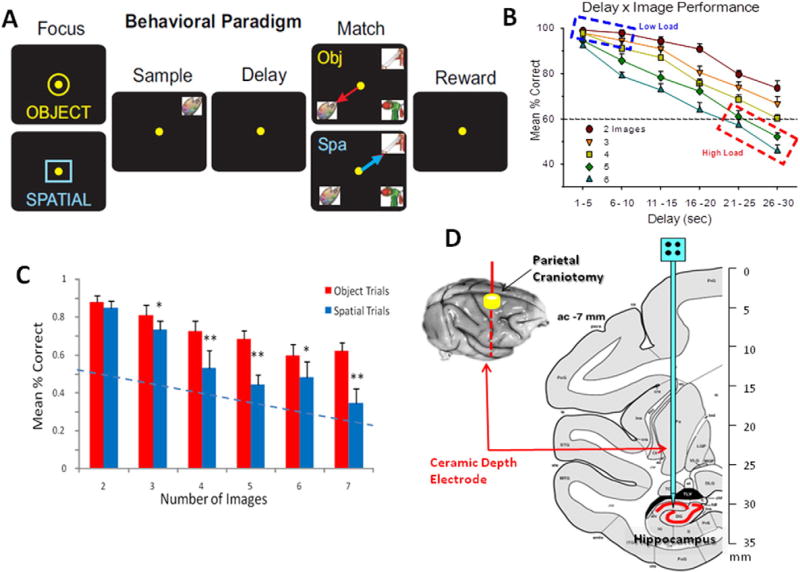
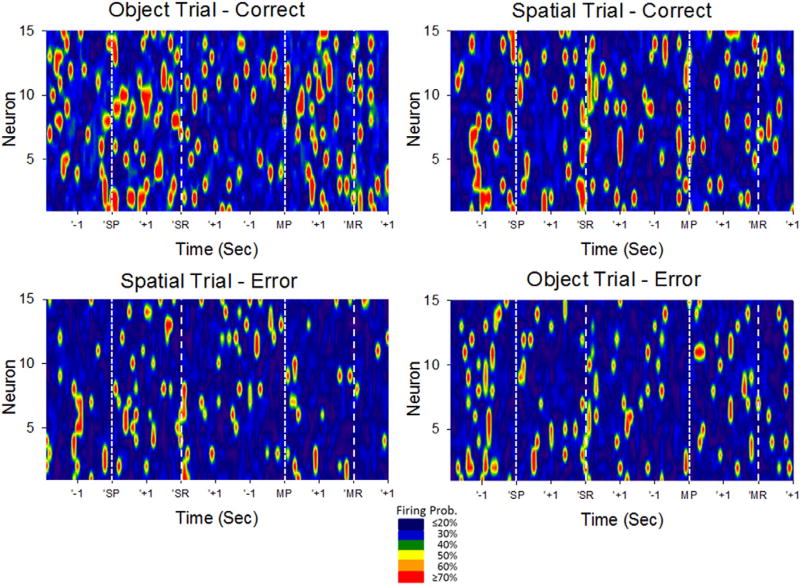
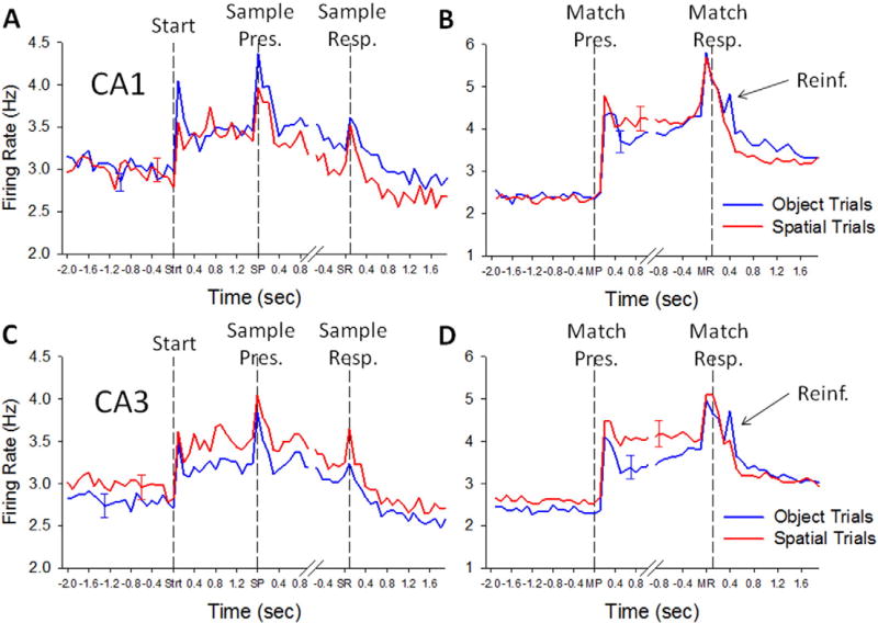
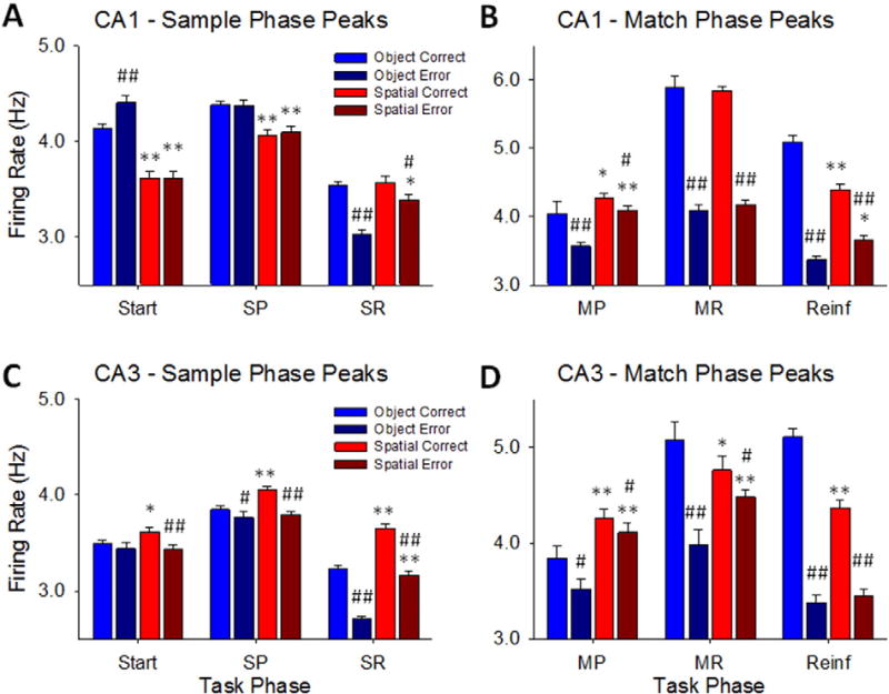
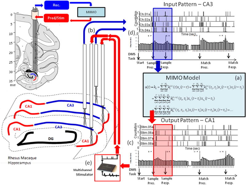
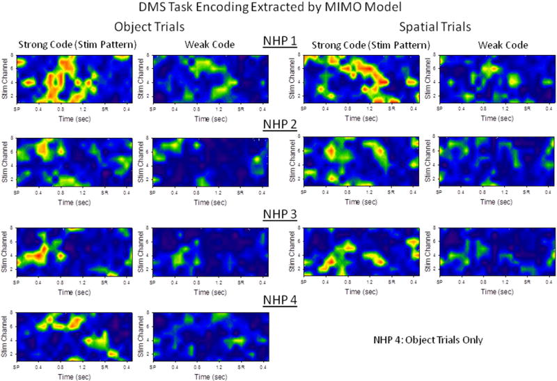
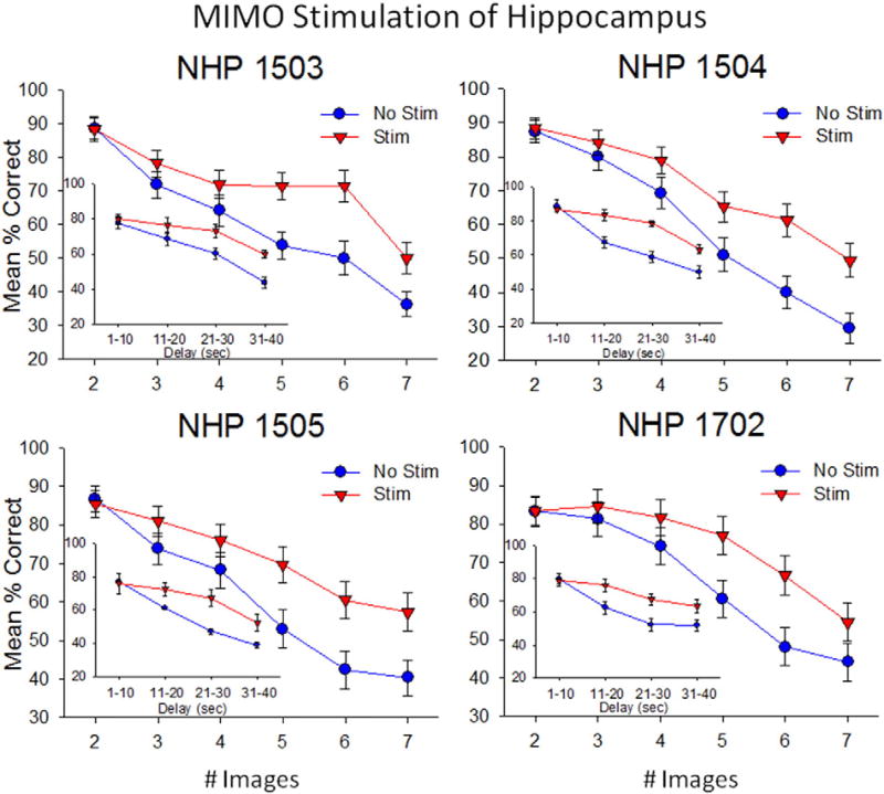
Very productive collaborative investigations characterized how multineuron hippocampal ensembles recorded in nonhuman primates (NHPs) encode short-term memory necessary for successful performance in a delayed match to sample (DMS) task and utilized that information to devise a unique nonlinear multi-input multi-output (MIMO) memory prosthesis device to enhance short-term memory in real-time during task performance. Investigations have characterized how the hippocampus in primate brain encodes information in a multi-item, rule-controlled, delayed match to sample (DMS) task. The MIMO model was applied via closed loop feedback micro-current stimulation during the task via conformal electrode arrays and enhanced performance of the complex memory requirements. These findings clearly indicate detection of a means by which the hippocampus encodes information and transmits this information to other brain regions involved in memory processing. By employing the nonlinear dynamic multi-input/multi-output (MIMO) model, developed and adapted to hippocampal neural ensemble firing patterns derived from simultaneous recorded multi-neuron CA1 and CA3 activity, it was possible to extract information encoded in the Sample phase of DMS trials that was necessary for successful performance in the subsequent Match phase of the task. The extension of this MIMO model to online delivery of electrical stimulation patterns to the same recording loci that exhibited successful CA1 firing in the DMS Sample Phase provided the means to increase task performance on a trial-by-trial basis. Increased utility of the MIMO model as a memory prosthesis was exhibited by the demonstration of cumulative increases in DMS task performance with repeated MIMO stimulation over many sessions. These results, reported below in this article, provide the necessary demonstrations to further the feasibility of the MIMO model as a memory prosthesis to recover and/or enhance encoding of cognitive information in humans with memory disruptions resulting from brain injury, disease or aging.

Keywords: Closed-loop; Electrical stimulation; Ensemble; Hippocampus; Memory; Neural prosthesis; Nonlinear model.


Figures

Fig. 1
Fig. 1
Illustration of DMS behavioral task and localization of hippocampal recording electrodes. A: Behavioral paradigm showing the sequence of events in the DMS task with correct cursor movement (yellow dot) indicated for each phase of the task: (Berger et al., 2005) Trial initiation ‘start signal’ (to maintain subject attention and signal the start of a new trial. Signal consists of yellow circle (upper) or blue square (lower) signaling an object or spatial trial, respectively. Placement of the cursor into the start signal initiated the Sample Phase of the trial. (Berger and Glanzman, 2005) Sample Presentation (SP) of a clip-art image in one of eight different spatial locations on the screen. The Sample Response (SR) consisted of movement of the cursor onto the presented sample image, which ended the Sample Phase and initiated the Delay Phase. (Chapin, 2004) Variable Delay consisted of randomly-selected 1–60 s interval with only a black screen showing. When computer determined that Delay interval had timed out, the Match Phase was initiated independent of any subject response. (Davachi, 2006) Match Presentation (MP) consisted of display of Sample image in a different location from Sample Phase, along with 1–7 Non-match distracter images. Match response (MR) consisted of cursor movements onto same image (for Object trials, red arrow) or same position (for Spatial trials, blue arrow) as in the Sample Phase. (Deadwyler et al., 1996) Correct MRs were rewarded by delivery of a squirt of juice reward (Reinf.). Placement of the cursor onto a non-match (distracter) image (object trial) or onto a different screen location from the SR (spatial trial) caused the screen to go blank without reward delivery. Inter-trial interval: 10.0 s. B: Overall performance averages showing the interaction of interposed delays with number of images presented in Match Phase. High and Low Cognitive Load conditions indicated by dashed outlines. C: Differential mean per cent correct performance in object and spatial trials (blue and red arrows in A) as a function of the number of (distracter) images presented in the match phase of the task. *p < 0.01, **p < 0.001 Object vs. Spatial. D: Diagram of NHP brain in cross-section showing hippocampal tetrode tracks through temporal lobe and placement in the CA3 and CA1 cell layers.
Fig. 2
Fig. 2
Ensemble encoding of DMS task events. Heat maps depict trial-based firing of hippocampal CA1 and CA3 neural ensembles averaged across 100 DMS trials. Vertical axis depicts individual cells (n = 15), while horizontal axis depicts elapsed time during typical DMS trials. DMS Sample Presentation (SP), Sample Response (SR), Match Presentation (MP) and Match Response (MR) are depicted on the horizontal axis by +1 and −1 s brackets around each event and by vertical dashed lines. Trials were sorted according to Object vs. Spatial trials, and correct vs. error performance prior to averaging firing rate for each cell within the session. Mean firing rate is normalized to probability of firing across trials, at a temporal resolution of 100 ms, and represented by the color code scale at the bottom. Data shown is from a single subject/single ensemble recording session.
Fig. 3
Fig. 3
Hippocampal CA1 and CA3 neuron firing in the Sample and Match Phases of the DMS task. A. Trial-based histograms (TBHs) of average firing of all CA1 cells (n = 431) recorded during Sample Phase from all 4 NHPs performing the DMS task. Each trace represents one of the 4 conditions listed on the left for comparison of correct Object vs. Spatial trials. The three events in the Sample phase (Start = Strt, Sample Present = SP, Sample Response = SR) are listed on the x-axis and marked by vertical dotted lines on each TBH. B. TBHs for Match Phase recorded from the same CA1 neurons as in A. Note the sustained elevated mean firing rates from Match onset until reward delivery or trial termination. Match Presentation (MP) and Match Response (MR) are indicated on the x-axis and by vertical dashed lines. The brief firing peaks that occurred coincident with reinforcement (Reinf.) occurred after the MR primarily on Object trials. C. TBHs of average firing for all CA3 cells (n = 801) recorded during Sample Phase. D. Match Phase TBHs for same CA3 neurons graphs as in C. Note that CA3 and CA1 neurons exhibited similar responses to the DMS trial events indicated. Error bars indicated peak S.E.M. for each TBH.
Fig. 4
Fig. 4
Comparison of Sample and Match Phase peak firing for Correct and Error Object and Spatial trials. A. Plots of CA1 mean (± S.E.M.) peak responses to the same 3 events in the Sample phase shown in Fig. 3A. Trials were sorted by Object and Spatial contingencies, for Correct vs. Error trials to show firing tendencies and differential encoding under different task conditions including correct and error trials. B. Mean (± S.E.M.) peak responses for CA1 neurons during two Match Phase events (MP, MR), plus Reinforcement (Reinf) delivery phase. C. Mean (± S.E.M.) peak Sample Phase responses (Start, SP, SR) for CA3 cells. D. Mean (± S.E.M.) peak Match Phase responses (MP, MR, Reinf) for CA3 cells. Start = Start Signal = trial initiation, SP = Sample Presentation, SR = Sample Response, MP = Match Presentation, MR = Match Response. *p < 0.01, **p < 0.001 Object vs. Spatial trial peaks, #p < 0.01, ##p < 0.001 Correct vs. Error trial peaks.
Fig. 5
Fig. 5
Multi-input, Multi-output (MIMO) nonlinear model used to: (a) calculate SR encoding via spatiotemporal firing relations between CA3 and CA1 recordings of neuron spike trains, (b) predict CA1 firing (c) fromCA3 recordings (d), and generate patterned stimulation (e) for feedback stimulation of the same CA1 areas. The anatomical diagrams show placement of CA3 and CA1 multi-cell recording tetrodes in the respective areas along the longitudinal axis of hippocampus. Recordings on correct DMS trials from these spatially distinct and layer specific tetrodes were fed into the MIMO model with CA3 as the input (blue arrow) and CA1 as the output pattern (red arrow). The MIMO model predicted correct CA1 output firing (i.e. “Strong Codes”) from CA3 inputs computed over the Sample Phase of the same trials (blue and red shaded rectangles) based on micro-temporal relationships between spike trains recorded at different spatial locations on correct trials. On stimulation trials, trains of electrical pulses mimicking the predicted Strong Code CA1 output spike trains were delivered to the same hippocampal electrode locations where the CA1 patterns were previously recorded. MIMO model stimulation patterns applied to the respective CA1 recording locations consisted of multi-channel biphasic pulses of 10–50 µA, 1.0 ms duration with a minimum 50 ms between pulses, at ≤20 stimulation pulses per second per channel. [Figure from (Hampson et al., 2013), used with permission]
Fig. 6
Fig. 6
Strong Sample Phase encoding extracted as a function of input-output relations of CA3 and CA1 firing on correct trials. The MIMO model was calculated using correct trials only for Object (left columns) and Spatial (right columns) trials, and yielded predictions of “Strong” (i.e. most likely to be correct) Sample codes for each of four NHPs tested (rows, NHP 1–4). Heat maps depict firing probability arranged as a spatio-temporal pattern of CA1 neurons (vertical axis) by time (horizontal axis) starting at Sample Presentation (SP) and extending for two seconds, including the occurrence of the Sample Response (SR). Strong code stimulation patterns (left side of each pair) were delivered whenever the MIMO-extracted CA1 firing was predicted to consist of a “Weak” code (right side of each pair). The Heat-map color shows a code for spike train firing probability (as predicted by MIMO model) ranging from <10% (dark blue) to ≥70% (red, see Fig. 2). Note that Weak code firing often shows lower probability of firing, but a similar spatio-temporal pattern to the Strong codes for the opposite trial type (i.e. Object vs. Spatial trial).
Fig. 7
Fig. 7
Facilitation of DMS task performance in four nonhuman primates (NHPs 1–4) following delivery of MIMO strong codes (Fig. 5b) via CA1 hippocampal stimulation derived from prior correct responses in previous sessions. Each graph shows the difference in performance for each of the 4 subjects on randomly selected stimulation trials vs. nonstimulated trials as a function of duration of the number (#) of distracter images (or intervening delay, insets). Strong code stimulation patterns (Stim) were delivered to CA1 hippocampal areas in the Sample phase on 40–50 of the different trial types during the same sessions in which 80–100 non-stimulated trials (No Stim) were also assessed on similar types of trials. Data in the above performance graphs were from 4 sessions per NHP subject. *p < 0.01, **p < 0.001 Stim vs. No Stim.

Similar articles

Cited by

References

    1. Berger TW, Glanzman DL. Toward Replacement Parts for the Brain. MIT Press; Cambridge, MA: 2005.
    1. Berger TW, Ahuja A, Courellis SH, Deadwyler SA, Erinjippurath G, Gerhardt GA, Gholmieh G, Granacki JJ, Hampson R, Hsaio MC, LaCoss J, Marmarelis VZ, Nasiatka P, Srinivasan V, Song D, Tanguay AR, Wills J. Restoring lost cognitive function. IEEE Eng Med. Biol. Mag. 2005;24:30–44. - PubMed
    1. Berger TW, Hampson RE, Song D, Goonawardena A, Marmarelis VZ, Deadwyler SA. A cortical neural prosthesis for restoring and enhancing memory. J. Neural Eng. 2011;8:046017. - PMC - PubMed
    1. Chapin JK. Using multi-neuron population recordings for neural prosthetics. Nat. Neurosci. 2004;7:452–455. - PubMed
    1. Davachi L. Item, context and relational episodic encoding in humans. Curr. Opin. Neurobiol. 2006;16:693–700. - PubMed

Publication types

MeSH terms