. 2016 Sep:93:215-25.
doi: 10.1016/j.nbd.2016.05.015. Epub 2016 May 24.

Suppression of glymphatic fluid transport in a mouse model of Alzheimer's disease

Affiliations

Suppression of glymphatic fluid transport in a mouse model of Alzheimer's disease

Weiguo Peng et al. Neurobiol Dis. 2016 Sep.

Abstract

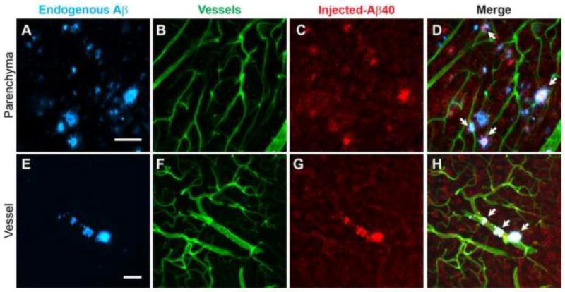
Glymphatic transport, defined as cerebrospinal fluid (CSF) peri-arterial inflow into brain, and interstitial fluid (ISF) clearance, is reduced in the aging brain. However, it is unclear whether glymphatic transport affects the distribution of soluble Aβ in Alzheimer's disease (AD). In wild type mice, we show that Aβ40 (fluorescently labeled Aβ40 or unlabeled Aβ40), was distributed from CSF to brain, via the peri-arterial space, and associated with neurons. In contrast, Aβ42 was mostly restricted to the peri-arterial space due mainly to its greater propensity to oligomerize when compared to Aβ40. Interestingly, pretreatment with Aβ40 in the CSF, but not Aβ42, reduced CSF transport into brain. In APP/PS1 mice, a model of AD, with and without extensive amyloid-β deposits, glymphatic transport was reduced, due to the accumulation of toxic Aβ species, such as soluble oligomers. CSF-derived Aβ40 co-localizes with existing endogenous vascular and parenchymal amyloid-β plaques, and thus, may contribute to the progression of both cerebral amyloid angiopathy and parenchymal Aβ accumulation. Importantly, glymphatic failure preceded significant amyloid-β deposits, and thus, may be an early biomarker of AD. By extension, restoring glymphatic inflow and ISF clearance are potential therapeutic targets to slow the onset and progression of AD.

Keywords: AQP4; Alzheimer's disease; Amyloid-β; Astrocytes; Brain ISF clearance; CSF; Clearance; Convective ISF flow; Glymphatic pathways; Lymphatic system.


Conflict of interest statement

Disclosure/Conflict of Interest. The authors declare no conflict of interest

Figures

Figure 1
Figure 1. Fluorescently-tagged Aβ40 and Aβ42 enter brain along the peri-arterial space and the pial membrane after intracisternal delivery
A) Schematic diagram showing the injection site. Representative images of Aβ40 and Aβ42 HiLyte Alexa 488 (B); cascade blue labeled dextran (CD-Dextran, a reference molecule; 10 kDa; C) and NG2-DsRed labeling of smooth muscle cells (D). E) Quantification of the areas of Aβ40, Aβ42 and CB-Dextran distribution. F) Standardization of Aβ distribution area as a percentage of CB-Dextran area. Peri-arterial inflow of CB-dextran and Aβ (G–J). Panel H shows the white boxed areas in G. White arrow head in panel H indicate the vessel magnified in panel I. J) Distribution of Aβ40 or Aβ42 (green) and CB-Dextran (blue) from an artery (red) shown in panel I. Vessel lumen in gray. Values are mean ± SEM, N=5. Scale bars: B, 2 mm; H, 500 μm; I, 100 μm. Aβ40-488 or Aβ42-488 and CD-dextran were intracisternally injected in NG2-DsRed reporter mice (2–3 months old) and the animals fixed with PFA 15 minutes later followed by preparation of vibratome sectioning and analysis of tracer distribution in brain sections.
Figure 2
Figure 2. Unlabeled human Aβ binds to cells after intracisternal delivery
A–D). Immunolabeling of CSF injected Aβ40 in brain (A), GFAP+-astrocytes (B),NeuN+-neurons (C) and merged images (D). E–H) Boxed area in panel A showing Aβ40 association with cells, mainly neurons (white arrows). I–L) Immunolabeling of Aβ42 (I), GFAP+-astrocytes (J), NeuN+-neurons (K) and merged images (L). M–P) Boxed area in panel I showing CSF Aβ42 mainly restricted to vessels. Representative images from 3 wild type mice (2–3 months old). Scale bars: A and I, 0.8 mm; E and M, 25 μm. Aβ40 or Aβ42 was intracisternally injected and after 30 minutes, to allow for cellular uptake, the brains were fixed and analyzed by immunolabeling.
Figure 3
Figure 3. Effect of Aβ on glymphatic inflow of CSF into brain
A) Schematic diagram of the experimental design. B) Representative images of vehicle (Veh) treated mice and mice treated with Aβ40 (C) and Aβ42 (D). Quantification of the distribution areas of Texas Red Dextran 3 kDa (E) and FITC-Dextran 40 kDa (F) from images as in panels B–D. CSF containing Aβ40 or Aβ42 (10 μM;5 μL), FITC-Dextran 40 kDa (1%) and Texas Red Dextran 3 kDa (1%) were intracisternally injected and after 30 min the PFA fixed brain sections were analyzed. Values are mean ± SEM, N=5–6 mice per group. G) Schematic diagram of the experimental design. Representative images of vehicle treated mice (H) and mice treated with Aβ40 (I) and Aβ42 (J). Quantification of the distribution areas of Texas Red Dextran 3 kDa (K) and FITC-Dextran40 kDa (L). First, 5 μL of CSF containing Aβ40 or Aβ42 (10 μM) was pre-infused intracisternally over 40 min, followed by CSF containing Aβ40 or Aβ42 (10 μM), FITC-Dextran40 kDa and Texas Red Dextran 3 kDa (1%), and the PFA fixed brain sections were analyzed. Values are mean ± SEM, N=6–9 mice (2–3 months old) per group.
Figure 4
Figure 4. Reduced CSF inflow and ISF clearance in APP/PS1 mice
A) Schematic diagram showing the injection site of the radiolabeled molecules. B–C) Inflow of 125I-Aβ40 and 14C-inulin from CSF into brain in young (3–4 months old; B) and old (C; 12–13 months old) APP/PS1mice (APP) compared to their controls (C). 125I-Aβ40 (10 nM) and 14C-inulin were injected intracisternally (1 μL/min for 5 min) and after 30 minutes the brain analyzed for radioactivity. N=4. D) Schematic diagram showing the cortical injection site. E–F) Clearance of 125I-Aβ40 and 14C-inulin in young (E; 6–8 months old) and old (F; 12–18 months old) APP/PS1mice compared to their controls. 125I-Aβ40 (10 nM) and 14C-inulin were microinjected intracortically (0.5 μL; 0.1 μL/min for 5 min) and after 30 minutes the brain analyzed for radioactivity. N=3–6. Values are mean ± SEM.
Figure 5
Figure 5. Intracisternal injected fluorescently labeled Aβ40 is associated with amyloid-β plaques in APP/PS1 mice
A–H) Endogenous Aβ detected with methoxy-X04 (A, E), cerebrovasculature delineated with intravascular injected lectin (B,F), CSF Aβ40-488 is present in brain (colored red) (C,G) and merged images (D,H) showing co-localization of the intracisternal injected Aβ40 with endogenous amyloid-β (white areas) in brain parenchyma and vessels. Representative images from 3 mice (7–9 months old). White areas show the co-localization of methoxy-X04 labeled endogenous amyloid-β plaques and injected Aβ40-488. Scale bars: A and E= 50 μm. Methoxy-X04 was administered intraperitoneally (IP; 10 mg/kg) and after 60 minutes Aβ40 was intracisternally injected, and the brain analyzed after an addition 30 minutes.
Figure 6
Figure 6. Levels of Aβ40, Aβ42 and oligomers are increased with age in the APP/PS1 mice
Levels of soluble Aβ40 (A) and Aβ42 (B), insoluble Aβ40 (C) and Aβ42 (D) and soluble Aβ oligomers (E) in cortical brain tissues of young (3–4 months old) and old (12–13 months old) mice. N=4–6 mice per group. Values are mean ± SEM.
Figure 7
Figure 7. Working model for the glymphatic distribution of Aβ
Aβ from the CSF enters brain (Aβ40>Aβ42) via the peri-arterial space and associated with parenchymal cells and amyloid β deposits. Aβ42 is mainly confined to the peri-arterial space due to, possibly, to the formation of fibrils, which is larger in size.

References

    1. Abbott NJ, et al. Astrocyte-endothelial interactions at the blood-brain barrier. Nat Rev Neurosci. 2006;7:41–53. - PubMed
    1. Banks WA, et al. Efflux of human and mouse amyloid beta proteins 1-40 and 1-42 from brain: impairment in a mouse model of Alzheimer’s disease. Neuroscience. 2003;121:487–92. - PMC - PubMed
    1. Bateman RJ, et al. Human amyloid-beta synthesis and clearance rates as measured in cerebrospinal fluid in vivo. Nat Med. 2006;12:856–61. - PMC - PubMed
    1. Bell RD, et al. Transport pathways for clearance of human Alzheimer’s amyloid beta-peptide and apolipoproteins E and J in the mouse central nervous system. J Cereb Blood Flow Metab. 2007;27:909–18. - PMC - PubMed
    1. Blennow K. Cerebrospinal fluid protein biomarkers for Alzheimer’s disease. NeuroRx. 2004;1:213–25. - PMC - PubMed

Publication types

Substances