Review
. 2016 May 27;14(1):33.
doi: 10.1186/s12969-016-0096-2.

Ultrasound in juvenile idiopathic arthritis

Affiliations
Review

Ultrasound in juvenile idiopathic arthritis

Silvia Magni-Manzoni. Pediatr Rheumatol Online J. .

Abstract

Background: In the recent years, musculoskeletal ultrasound (MSUS) has been regarded as especially promising in the assessment of juvenile idiopathic arthritis (JIA), as a reliable method to precisely document and monitor the synovial inflammation process.

Main content: MSUS is particularly suited for examination of joints in children due to several advantages over other imaging modalities. Some challenges should be considered for correct interpretation of MSUS findings in children, due to the peculiar features of the growing skeleton. MSUS in JIA is considered particularly useful for its ability to detect subclinical synovitis, to improve the classification of patients in JIA subtypes, for the definition of remission, as guidance to intraarticular corticosteroid injections and for capturing early articular damage. Current evidence and applications of MSUS in JIA are documented by several authors. Recent advances and insights into further investigations on MSUS in healthy children and in JIA patients are presented and discussed in the present review.

Conclusions: MSUS shows great promise in the assessment and management of children with JIA. Nonetheless, anatomical knowledge of sonographic changes over time, underlying immunopathophysiology, standardization and validation of MSUS in healthy children and in patients with JIA are still under investigation. Further research and educational efforts are required for expanding this imaging modality to more clinicians in their daily practice.

Keywords: Children; Juvenile idiopathic arthritis; Musculoskeletal ultrasound; Pediatric rheumatology.


Figures
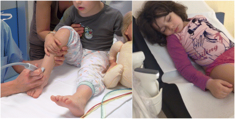
Fig. 1
Fig. 1
MSUS is well accepted by children and their parents, and can be performed as completion of the joint examination during the standard clinical assessment of the child. Some children have fun during MSUS evaluation, others totally relax and can fall asleep
Fig. 2
Fig. 2
Longitudinal scan of the suprapatellar recess in two 7 years old boys performed with high multifrequency probe: a. Top-level ultrasound machine in the early 2000’s; b. Top-level ultrasound machine in 2015
Fig. 3
Fig. 3
Metaphysis (M) look like erosions; epiphysis (E) and unossified bones are anechoic, like synovial effusion (a,b). Physiological vascularization at insertion of enthesis to the cartilage can be frequently detected, resembling enthesitis (c). Feeding vessels can be intraarticular or close to ossifying nuclei, and must not be considered as signs of active synovitis (d). a. Dorsal longitudinal scan of the II metacarpophalangeal joint in a 2 years old child, grey-scale. b. Longitudinal medial scan of the suprapatellar recess in a 2 years old child, grey-scale. c. Longitudinal scan of the Achilles tendon at insertion to the calcaneus in a 5 years old child, power Doppler d. Longitudinal medial scan of the suprapatellar recess in a 5 years old child, power Doppler
Fig. 4
Fig. 4
A 10 years old girl with JIA presented with mild swelling of the left ankle, without tenderness/pain on motion or limitation on motion. MSUS showed no signs of synovitis at the tibiotalar joint (a), and allowed detection of a hypo-anechoic halo around the medial tendons (b) and pathologic vascularization on power Doppler along both the posterior tibialis tendon (c) and the common flexor digiti tendon (d), indicating flourishing tenosynovitis. PTT: posterior tibialis tendon; CDT: common flexor digitorum tendon; A: posterior tibialis artery; V: posterior tibialis vein
Fig. 5
Fig. 5
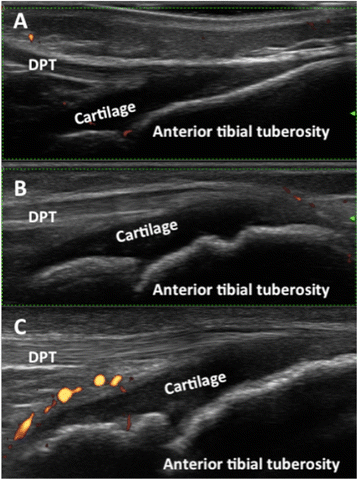
Sonographic appearance of the patellar enthesis at different ages. a. 2 years old girl; b. 8 years old girl; c. 10 years old boy. DPT: distal patellar tendon
Fig. 6
Fig. 6
MSUS allows detection of the exact location of inflammation and correct needle placement, even in very small sites, such as the synovial sheath of the flexor tendon of the VI digit in a 3 years old girl. NT: needle tip; FDT: flexor digiti tendon; SE: synovial effusion
Fig. 7
Fig. 7
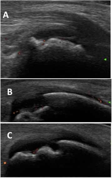
The calcaneus bone appears fragmented in children respectively aged 6 years (a), 8 years (b), and 10 years (c). Feeding vessels can be detected by power Doppler (red dots)

References

    1. Ravelli A, Martini A. Juvenile idiopathic arthritis. Lancet. 2007;369:767–78. doi: 10.1016/S0140-6736(07)60363-8. - DOI - PubMed
    1. Kessler EA, Becker ML. Therapeutic advancements in juvenile idiopathic arthritis. Best Pract Res Clin Rheumatol. 2014;28:293–313. doi: 10.1016/j.berh.2014.03.005. - DOI - PubMed
    1. Webb K, Wedderburn LR. Advances in the treatment of polyarticular juvenile idiopathic arthritis. Curr Opin Rheumatol. 2015;27:505–10. doi: 10.1097/BOR.0000000000000206. - DOI - PMC - PubMed
    1. Hinze C, Gohar F, Foell D. Management of juvenile idiopathic arthritis: hitting the target. Nat Rev Rheumatol. 2015;11:290–300. doi: 10.1038/nrrheum.2014.212. - DOI - PubMed
    1. Naredo E, Wakefield RJ, Iagnocco A, et al. The OMERACT ultrasound task force--status and perspectives. J Rheumatol. 2011;38:2063–7. doi: 10.3899/jrheum.110425. - DOI - PubMed

MeSH terms

Substances