

Microsurgical resection of tumors of the lateral and third ventricles: operative corridors for difficult-to-reach lesions
- PMID: 27235145
- PMCID: PMC5090015
- DOI: 10.1007/s11060-016-2126-9
Microsurgical resection of tumors of the lateral and third ventricles: operative corridors for difficult-to-reach lesions
Abstract
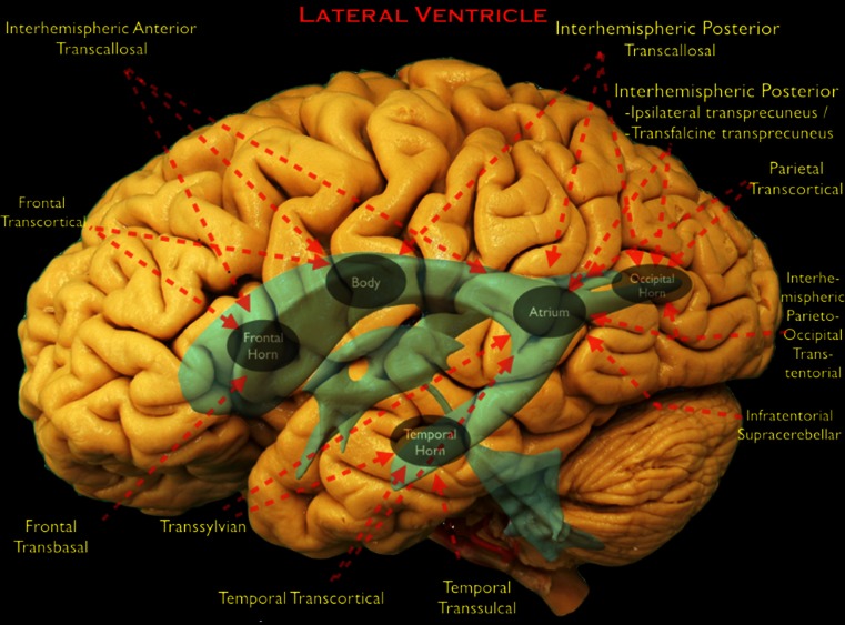
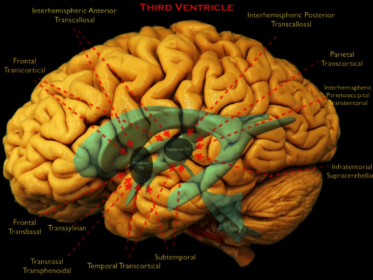
Tumors of the lateral and third ventricles are cradled on all sides by vital vascular and eloquent neural structures. Microsurgical resection, which always requires attentive planning, plays a critical role in the contemporary management of these lesions. This article provides an overview of the open microsurgical approaches to the region highlighting key clinical perspectives.
Keywords: Brain tumor surgery; Lateral ventricle; Microneurosurgery; Surgical approach; Third ventricle.
Conflict of interest statement
Dr. Aaron Cohen–Gadol has a consulting agreement with Zeiss Meditec, the rest of the authors declare that they have no conflict of interest. Informed consent Informed consent was obtained from all individual participants included in the study.
Figures
Similar articles
-
Transcallosal approach to third ventricle tumors: how I do it.Acta Neurochir (Wien). 2013 Jun;155(6):1031-4. doi: 10.1007/s00701-013-1714-0. Epub 2013 Apr 26. Acta Neurochir (Wien). 2013. PMID: 23619958
-
The use of transcallosal-interforniceal approach for microsurgical removal of the third ventricle tumors.J Neurosurg Sci. 2015 Mar;59(1):19-24. Epub 2014 Oct 8. J Neurosurg Sci. 2015. PMID: 25294411
-
Primary tumors of the lateral ventricles of the brain.Chirurgia (Bucur). 2013 Sep-Oct;108(5):616-30. Chirurgia (Bucur). 2013. PMID: 24157104
-
Surgery of intraventricular tumors.Neurosurgery. 2008 Jun;62(6 Suppl 3):1029-40; discussion 1040-1. doi: 10.1227/01.neu.0000333768.12951.9a. Neurosurgery. 2008. PMID: 18695523 Review.
-
[Ependymomas of the lateral ventricle. A series of 27 cases with review of the literature].Neurochirurgie. 2011 Sep-Dec;57(4-6):206-9. doi: 10.1016/j.neuchi.2011.09.022. Epub 2011 Oct 24. Neurochirurgie. 2011. PMID: 22030173 Review. French.
Cited by
-
Discernible interindividual patterns of global efficiency decline during theoretical brain surgery.Sci Rep. 2024 Jun 25;14(1):14573. doi: 10.1038/s41598-024-64845-4. Sci Rep. 2024. PMID: 38914649 Free PMC article.
-
Effect comparison of neuroendoscopy versus microsurgery in the treatment of lateral ventricular tumors.Front Oncol. 2023 Jul 24;13:1008291. doi: 10.3389/fonc.2023.1008291. eCollection 2023. Front Oncol. 2023. PMID: 37554163 Free PMC article.
-
Endoscopic transventricular approach for the resection of a hemorrhagic cavernous malformation of the tectal plate: Operative video.Surg Neurol Int. 2023 Feb 3;14:45. doi: 10.25259/SNI_57_2023. eCollection 2023. Surg Neurol Int. 2023. PMID: 36895228 Free PMC article.
-
Endoscopic management of a cavernous malformation on the floor of third ventricle and aqueduct of Sylvius: Technical case report and review of the literature.Surg Neurol Int. 2017 Sep 26;8:237. doi: 10.4103/sni.sni_165_17. eCollection 2017. Surg Neurol Int. 2017. PMID: 29026673 Free PMC article.
-
Intraventricular neuroepithelial tumors: surgical outcome, technical considerations and review of literature.BMC Cancer. 2020 Nov 3;20(1):1060. doi: 10.1186/s12885-020-07570-1. BMC Cancer. 2020. PMID: 33143683 Free PMC article.
References
Publication types
MeSH terms
LinkOut - more resources
Full Text Sources
Other Literature Sources
