. 2016 Jul 29;291(31):16100-11.
doi: 10.1074/jbc.M116.724815. Epub 2016 May 27.

Distal Hydrogen-bonding Interactions in Ligand Sensing and Signaling by Mycobacterium tuberculosis DosS

Affiliations

Distal Hydrogen-bonding Interactions in Ligand Sensing and Signaling by Mycobacterium tuberculosis DosS

Debashree Basudhar et al. J Biol Chem. .

Abstract

Mycobacterium tuberculosis DosS is critical for the induction of M. tuberculosis dormancy genes in response to nitric oxide (NO), carbon monoxide (CO), or hypoxia. These environmental stimuli, which are sensed by the DosS heme group, result in autophosphorylation of a DosS His residue, followed by phosphotransfer to an Asp residue of the response regulator DosR. To clarify the mechanism of gaseous ligand recognition and signaling, we investigated the hydrogen-bonding interactions of the iron-bound CO and NO ligands by site-directed mutagenesis of Glu-87 and His-89. Autophosphorylation assays and molecular dynamics simulations suggest that Glu-87 has an important role in ligand recognition, whereas His-89 is essential for signal transduction to the kinase domain, a process for which Arg-204 is important. Mutation of Glu-87 to Ala or Gly rendered the protein constitutively active as a kinase, but with lower autophosphorylation activity than the wild-type in the Fe(II) and the Fe(II)-CO states, whereas the E87D mutant had little kinase activity except for the Fe(II)-NO complex. The H89R mutant exhibited attenuated autophosphorylation activity, although the H89A and R204A mutants were inactive as kinases, emphasizing the importance of these residues in communication to the kinase core. Resonance Raman spectroscopy of the wild-type and H89A mutant indicates the mutation does not alter the heme coordination number, spin state, or porphyrin deformation state, but it suggests that interdomain interactions are disrupted by the mutation. Overall, these results confirm the importance of the distal hydrogen-bonding network in ligand recognition and communication to the kinase domain and reveal the sensitivity of the system to subtle differences in the binding of gaseous ligands.

Keywords: Mycobacterium tuberculosis; biosensor; carbon monoxide; heme; hydrogen bonding network; molecular dynamics; nitric oxide; oxygen binding; resonance Raman.


Figures

FIGURE 1.
FIGURE 1.
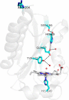
Crystal structure of the ferric DosS GAF-A(63–210) domain (Protein Data Bank code 2W3E) showing the proximal histidine ligand and the distal hydrogen-bonding network. A hydrogen bond between Arg-204 and His-89 is not evident in the crystal structure but is observed in the molecular dynamics simulations.
FIGURE 2.
FIGURE 2.
UV-visible spectrophotometric characterization of the H89R (A), H89A (B), E87D (C), E87G (D), E87A (E), and R204A (F) mutants in the Fe(II) unligated state (red) and coordinated with CO (black), NO (green), and after exposure to O2 (magenta). This latter condition in most cases resulted in complete or nearly complete autoxidation to the Fe(III) state.
FIGURE 3.
FIGURE 3.
High frequency resonance Raman spectra of ferric and ferrous wild-type and H89A variant proteins obtained with a 413-nm excitation (A) and low frequency resonance Raman spectra of ferrous wild-type and H89A variant proteins obtained with a 442-nm excitation (B).
FIGURE 4.
FIGURE 4.
Low frequency 16O2-18O2 resonance Raman difference spectra of the oxy complexes of full-length wild-type (blue trace), GAF-A wild-type domain (black trace), and full-length H89A (red trace) DosS proteins (A); high frequency spectra of the carbonyl complexes formed with 12CO, 13CO, and the resulting 12CO-13CO difference spectra (B), and 14N16O-15N18O difference spectra of the nitrosyl complexes (C) of full-length wild-type (blue traces), GAF-A wild-type domain (black traces), and full-length H89A (red traces) DosS proteins. All spectra were obtained at room temperature with a 413-nm excitation.
FIGURE 5.
FIGURE 5.
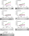
Representative autophosphorylation gel and quantification of the activity of wild-type (A), H89R (B), H89A (C), E87D (D), E87G (E), E87A (F), and R204A (G) mutants of DosS in the Fe(II) state (red) and after its exposure to CO (black), O2 (magenta), and NO (green). 1st lanes, wild-type bound to CO at 20 min; 2nd to 5th lanes, Fe(II); 6th to 9th lanes, exposure of Fe(II) to CO; 10th to 13 lanes, exposure of Fe(II) to O2; and 13th to 17th lanes, exposure of Fe(II) to NO at 3-, 6-, 9-, and 12-min time points, respectively.
FIGURE 6.
FIGURE 6.
Most frequently observed hydrogen-bonding patterns observed in molecular dynamics simulations for the wild-type DosS GAF-A domain and its E87A and E87G mutants.
FIGURE 7.
FIGURE 7.
Graphic depiction of the hydrogen-bonding patterns observed as a function of time in the molecular dynamics simulations of the wild-type DosS GAF-A (four upper traces) and the E87A mutant (two lower traces). The open circles indicate the presence of the indicated hydrogen-bonding pattern at a given time point in the simulation. The legend on the left indicates the H-bonding pattern.
FIGURE 8.
FIGURE 8.
Comparison of the local water network in the wild-type ferric enzyme (A) with that of the ferrous enzyme (B). Note the difference in the positions of Glu-87 in the two structures as indicated by the orange discs.
FIGURE 9.
FIGURE 9.
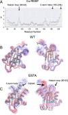
A, RMSF plots over the course of the molecular dynamics simulation for the wild-type (green), E87A (blue), and E87G (orange) mutants. To aid in visualizing this fluctuation, an amalgamation of snapshots (every 100 frames) are overlaid in (B and C) for the wild-type and E87A mutants, with each shown in two orientations reflecting a 90° rotation about a vertical axis. The snapshots are color-coded chronologically, with the earliest in darkest blue and the latest in darkest red. The wild-type has considerably less movement than the E87A mutant.
FIGURE 10.
FIGURE 10.
Sequence alignment of a region of M. tuberculosis DosS and DosT with similar regions from other organisms obtained using Clustal Omega via the web interface, where the sequences are from M. tuberculosis DosS, M. tuberculosis DosT, Mycobacterium bovis A0A0H3P5J6, Mycobacterium marinum B2HGI2, and Mycobacterium ulcerans A0PR05. The numbering indicated at top is from M. tuberculosis DosS.

References

    1. Dye C., Scheele S., Dolin P., Pathania V., and Raviglione M. C. (1999) Consensus statement. Global burden of tuberculosis: estimated incidence, prevalence, and mortality by country. WHO Global Surveillance and Monitoring Project. J. Am. Med. Assoc. 282, 677–686 - PubMed
    1. Parrish N. M., Dick J. D., and Bishai W. R. (1998) Mechanisms of latency in Mycobacterium tuberculosis. Trends Microbiol. 6, 107–112 - PubMed
    1. Fenton M. J., Vermeulen M. W., Kim S., Burdick M., Strieter R. M., and Kornfeld H. (1997) Induction of γ interferon production in human alveolar macrophages by Mycobacterium tuberculosis. Infect. Immun. 65, 5149–5156 - PMC - PubMed
    1. Silva-Gomes S., Appelberg R., Larsen R., Soares M. P., and Gomes M. S. (2013) Heme catabolism by heme oxygenase-1 confers host resistance to Mycobacterium infection. Infect. Immun. 81, 2536–2545 - PMC - PubMed
    1. Regev D., Surolia R., Karki S., Zolak J., Montes-Worboys A., Oliva O., Guroji P., Saini V., Steyn A. J., Agarwal A., and Antony V. B. (2012) Heme oxygenase-1 promotes granuloma development and protects against dissemination of mycobacteria. Lab. Invest. 92, 1541–1552 - PMC - PubMed

Publication types

MeSH terms