. 2016 Jul 22;291(30):15867-80.
doi: 10.1074/jbc.M116.727008. Epub 2016 May 27.

Regulation of Connexin43 Function and Expression by Tyrosine Kinase 2

Affiliations

Regulation of Connexin43 Function and Expression by Tyrosine Kinase 2

Hanjun Li et al. J Biol Chem. .

Abstract

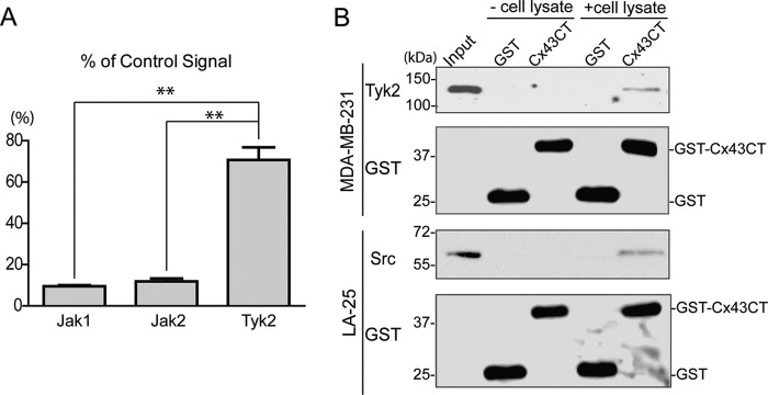
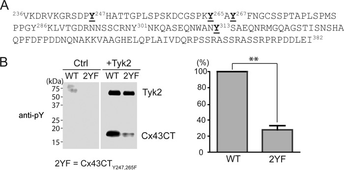
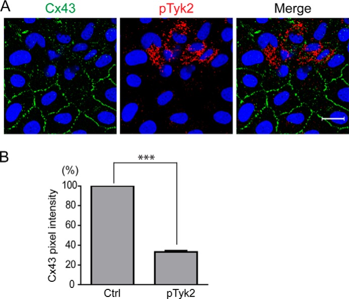
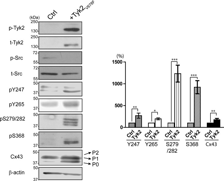
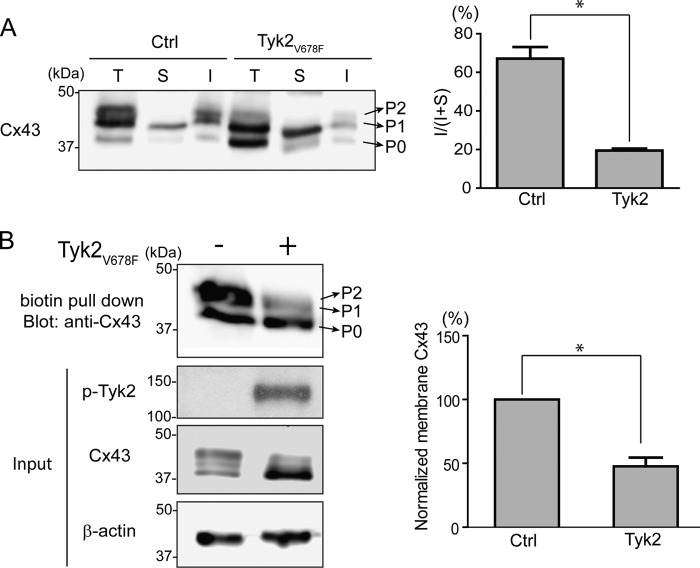
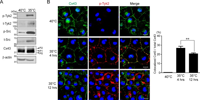
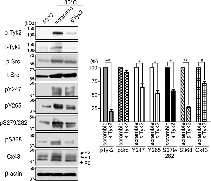
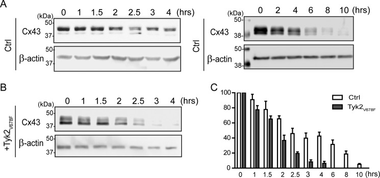
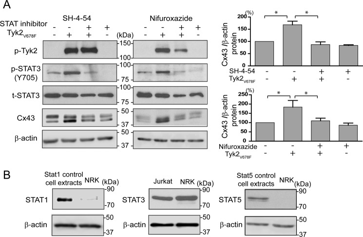
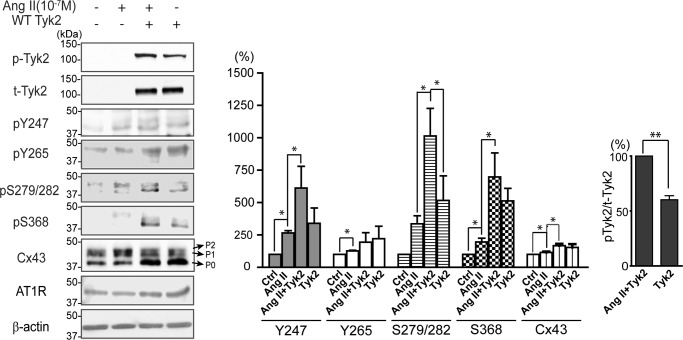
Connexin43 (Cx43) assembly and degradation, the regulation of electrical and metabolic coupling, as well as modulating the interaction with other proteins, involve phosphorylation. Here, we identified and characterized the biological significance of a novel tyrosine kinase that phosphorylates Cx43, tyrosine kinase 2 (Tyk2). Activation of Tyk2 led to a decrease in Cx43 gap junction communication by increasing the turnover rate of Cx43 from the plasma membrane. Tyk2 directly phosphorylated Cx43 residues Tyr-247 and Tyr-265, leading to indirect phosphorylation on residues Ser-279/Ser-282 (MAPK) and Ser-368 (PKC). Although this phosphorylation pattern is similar to what has been observed following Src activation, the response caused by Tyk2 occurred when Src was inactive in NRK cells. Knockdown of Tyk2 at the permissive temperature (active v-Src) in LA-25 cells decreased Cx43 phosphorylation, indicating that although activation of Tyk2 and v-Src leads to phosphorylation of the same Cx43CT residues, they are not identical in level at each site. Additionally, angiotensin II activation of Tyk2 increased the intracellular protein level of Cx43 via STAT3. These findings indicate that, like Src, Tyk2 can also inhibit gap junction communication by phosphorylating Cx43.

Keywords: Western blot; cell biology; cell signaling; gap junction; phosphorylation.


Figures

FIGURE 1.
FIGURE 1.
Phosphorylation of the Cx43CT domain by JAK tyrosine kinases. A, an in vitro kinase assay was performed using the catalytic domain of Jak1, Jak2, and Tyk2 to phosphorylate purified Cx43CT. The amount of Cx43CT phosphorylation was compared with a positive control peptide for each kinase (100% signal) (**, p < 0.01). B, purified GST (26 kDa) or GST-Cx43CT (42 kDa) bound on glutathione-agarose beads was incubated with or without MDA-MB-231 cell lysate (top), and the pulled down product was analyzed by Western blotting analysis using an anti-Tyk2 antibody. GST-Cx43CT pulldown of Src from LA-25 cell lysate was used as the positive control (bottom).
FIGURE 2.
FIGURE 2.
Identification of the Cx43CT tyrosine residues phosphorylated by Tyk2. A, sequence of the Cx43CT domain. The Cx43CT tyrosine residues identified from mass spectrometry to be phosphorylated by the Tyk2 catalytic domain in vitro are highlighted (bold and underlined). B, the same in vitro kinase assay as described in A was performed using wild-type Cx43CT236–382 or a Y247,265F (2YF) mutant as substrate, and phosphorylation was detected by Western blotting using a general anti-phosphotyrosine antibody. The control (Ctrl) group did not contain Tyk2. The phosphotyrosine level was quantified using ImageJ software (n = 3, **, p < 0.01).
FIGURE 3.
FIGURE 3.
Effect of Tyk2 on the cellular localization of Cx43. A, cellular localization of endogenous Cx43 and constitutively active Tyk2 (Tyk2V678F) in NRK cells detected by immunofluorescence (green, Cx43; blue, DAPI-stained DNA; red, active Tyk2). Scale bar is 20 μm. B, quantification of Cx43 expression level at the plasma membrane. Cx43 pixel intensity of 204 cell pairs containing p-Tyk2 was normalized to the Cx43 pixel intensity of 204 cell pairs without p-Tyk2 (Ctrl) by ImageJ software. Cell pairs with or without p-Tyk2 were from the same images. The data are representative of three independent experiments (***, p < 0.001).
FIGURE 4.
FIGURE 4.
Phosphorylation of Cx43 by Tyk2 in NRK cells. Western blotting of active Tyk2 (p-Tyk2), total Tyk2, active Src (p-Src), total Src, Cx43 pTyr-247, pTyr-265, pSer-279/pSer-282, pSer-368, and total Cx43 from NRK cells without (Ctrl, control) or with transfection of Tyk2V678F. The Cx43 mobility shifts (P0, P1, and P2) are labeled. Relative protein levels were quantified by analyzing the scanned blots using ImageJ software with normalization of protein expression to the control lane (value set arbitrarily as 100%). The data are representative of three independent experiments (*, p < 0.05; **, p < 0.01; ***, p < 0.001).
FIGURE 5.
FIGURE 5.
Effect of Tyk2 on the plasma membrane localization of Cx43 in NRK cells. A, Triton X-100 solubility assay. Equal amounts of total protein fraction (T), Triton X-100-soluble fraction (S), and the insoluble fraction (I) were run on SDS-PAGE and blotted with anti-Cx43 antibody. Protein levels were quantified to determine the insoluble to insoluble + soluble ratio [I/(I + S)] (*, p < 0.05). B, biotinylation assay. NRK cells with or without Tyk2V678F transfection were cell surface-biotinylated. Biotinylated proteins were pulled down by immobilization on streptavidin-agarose beads and immunoblotted for Cx43. Protein levels of biotinylated Cx43 were quantified (*, p < 0.05). Input shows active Tyk2 and Cx43 protein in the cell lysate. Ctrl, control.
FIGURE 6.
FIGURE 6.
Colocalization of endogenous Tyk2 and Cx43 in LA-25 cells. A, Western blotting of active Tyk2 (p-Tyk2), total Tyk2, Src (p-Src), total Src and Cx43 in LA-25 cells at 40 and 35 °C. B, cellular localization of endogenous Cx43 and active Tyk2 in LA-25 cells at 40 °C or 35 °C was visualized by using immunofluorescence (green, Cx43; blue, DAPI-stained DNA; red, Tyk2). Scale bar is 20 μm. Colocalization of Cx43 and p-Tyk2 was analyzed based on 12 images from three independent experiments. The Manders method was used to measure the green signal (Cx43) coincident with the red signal (active Tyk2) over the total intensity of green signal (**, p < 0.01).
FIGURE 7.
FIGURE 7.
Effect of Tyk2 knockdown on the phosphorylation level of Cx43 in LA-25 cells. Western blotting of active Tyk2 (p-Tyk2), total Tyk2, active Src (p-Src), total Src, Cx43 pTyr-247, pTyr-265, pSer-279/pSer-282, pSer-368, and total Cx43 from LA-25 cells at 40 or 35 °C treated with scrambled or Tyk2 siRNA for 12 h. Relative protein levels were quantified using ImageJ software and normalized to the expression level in the scramble RNA-treated sample at 35 °C (*, p < 0.05; **, p < 0.01).
FIGURE 8.
FIGURE 8.
Effect of Tyk2 on the turnover rate of Cx43 in NRK cells. Control (A, Ctrl) or transfected NRK cells (B) with the constitutively active Tyk2 (Tyk2V678F) construct were treated with 100 μg/ml cycloheximide for different durations prior to lysis. Total Cx43 protein was immunoblotted. C, the protein level of three independent experiments was quantified using ImageJ software and normalized to the protein level at 0 h.
FIGURE 9.
FIGURE 9.
Effect of STAT3 activation by Tyk2 on Cx43 mRNA level. A, RT-PCR shows Cx43 mRNA level with or without transfection of active Tyk2 (Tyk2V678F). The appropriate number of cycles was determined by testing different number of cycles (16, 21, 24, 27, and 30 cycles) for Cx43 amplification (top panel). Cx43 intensity increased up to 24 cycles, where a plateau was reached; thus 24 cycles were used to run semiquantitative RT-PCR. Cx43 and β-actin were amplified for 24 cycles and ran on 2% agarose gel (right panel). B, NRK cells with or without active Tyk2 (Tyk2V678F) transfection for 8 h were treated with the STAT3/5 inhibitor SH-4-54 (5 μm) or the STAT1/3/5 inhibitor nifuroxazide (50 μm) for another 16 h. Cx43 and β-actin were amplified for 24 cycles and run on 2% agarose gel. Cx43 and β-actin mRNA levels were quantified by densitometry from three independent experiments (*, p < 0.05; **, p < 0.01).
FIGURE 10.
FIGURE 10.
Effect of STAT3 activation by Tyk2 on Cx43 protein level. A, Western blotting analysis of active Tyk2 (p-Tyk2), active STAT3 (p-STAT3), total STAT3, and Cx43 in NRK cells transfected with or without active Tyk2 (Tyk2V678F) and treated with and without SH-4-54 or nifuroxazide. Cx43 protein level was quantified using ImageJ software (*, p < 0.05). B, Western blotting analysis of STAT1, STAT3, and STAT5 from NRK cell lysate. Cell extracts from HeLa cells treated with IFN-α (100 ng/ml) for 5 min were used as a positive control for blotting STAT1 and STAT5. Jurkat cell lysate was used as the positive control for blotting STAT3.
FIGURE 11.
FIGURE 11.
Effect of Ang II activation of Tyk2 on the phosphorylation level of Cx43. Western blotting analysis of active Tyk2 (p-Tyk2), total Tyk2, pTyr-247, Cx43 pTyr-265, pSer-279/pSer-282, pSer-368, total Cx43, and the angiotensin II type 1 receptor (AT1R) from NRK cells with or without transfected WT Tyk2 and treated with or without Ang II (10−7 m). The protein phosphorylation levels were quantified using ImageJ software (*, p < 0.05; **, p < 0.01).
FIGURE 12.
FIGURE 12.
Effect of Cx43 Tyr-247 phosphorylation on the interaction with β-tubulin. Western blotting analysis of β-tubulin from a biotin pulldown assay using a biotinylated-phosphopeptide, Cx43CT-(234–255) (pY247) or non-phosphopeptide (Y247nop) bound to streptavidin beads and incubated with NRK cell lysate. Biotin bound to streptavidin-agarose beads was used as the negative control. Protein levels were quantified using ImageJ (*, p < 0.05) and normalized by input (1:20 loaded compared with pulldown groups) from three independent experiments.

References

    1. Laird D. W. (2006) Life cycle of connexins in health and disease. Biochem. J. 394, 527–543 - PMC - PubMed
    1. Solan J. L., and Lampe P. D. (2005) Connexin phosphorylation as a regulatory event linked to gap junction channel assembly. Biochim. Biophys. Acta 1711, 154–163 - PubMed
    1. Solan J. L., and Lampe P. D. (2007) Key connexin 43 phosphorylation events regulate the gap junction life cycle. J. Membr. Biol. 217, 35–41 - PMC - PubMed
    1. Axelsen L. N., Calloe K., Holstein-Rathlou N. H., and Nielsen M. S. (2013) Managing the complexity of communication: regulation of gap junctions by post-translational modification. Front. Pharmacol. 4, 130. - PMC - PubMed
    1. Lampe P. D., and Lau A. F. (2004) The effects of connexin phosphorylation on gap junctional communication. Int. J. Biochem. Cell Biol. 36, 1171–1186 - PMC - PubMed

LinkOut - more resources