

IL-2, IL-4, IFN-γ or TNF-α enhances BAFF-stimulated cell viability and survival by activating Erk1/2 and S6K1 pathways in neoplastic B-lymphoid cells
- PMID: 27235588
- PMCID: PMC4905560
- DOI: 10.1016/j.cyto.2016.05.017
IL-2, IL-4, IFN-γ or TNF-α enhances BAFF-stimulated cell viability and survival by activating Erk1/2 and S6K1 pathways in neoplastic B-lymphoid cells
Abstract
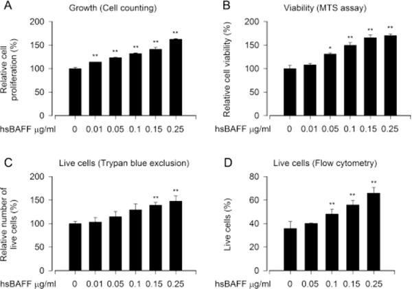
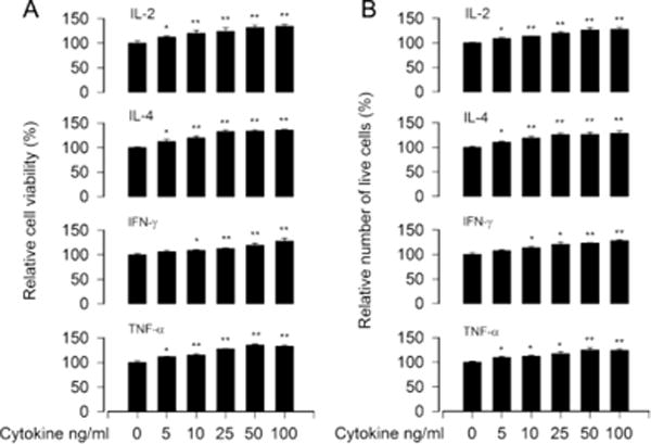
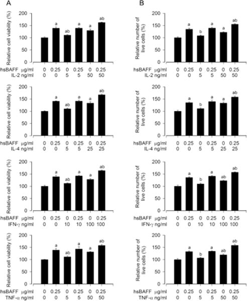
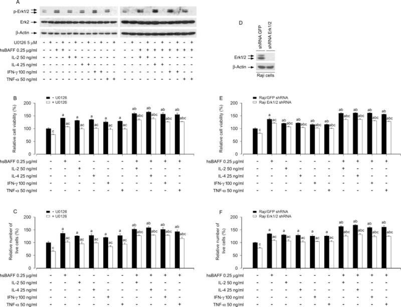
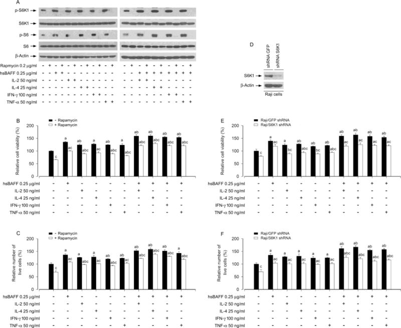
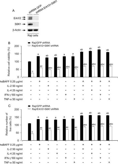
B-cell activating factor of the TNF family (BAFF) has been documented to act as a critical factor in the development of aggressive B lymphocytes and autoimmune diseases. However, the effect of various cytokines on BAFF-elicited neoplastic B-lymphoid cells is not known. In this study, we exhibited that administration of human soluble BAFF (hsBAFF), IL-2, IL-4, IFN-γ, or TNF-α alone increased cell viability and survival in Raji cells concentration-dependently, yet a more robust viability/survival was seen in the cells co-treatment of IL-2, IL-4, IFN-γ, or TNF-α with hsBAFF, respectively. Further research revealed that both Erk1/2 and S6K1 signaling pathways were essential for IL-2, IL-4, IFN-γ, or TNF-α enhancement of the viability/survival in the hsBAFF-stimulated cells, as inhibition of Erk1/2 with U0126 or down-regulation of Erk1/2, or blockage of S6K1 with rapamycin or silencing S6K1, or silencing S6K1/Erk1/2, respectively, reduced the cell viability/survival in the cells treated with/without hsBAFF±IL-2, IL-4, IFN-γ, or TNF-α. These findings indicate that IL-2, IL-4, IFN-γ or TNF-α enhances BAFF-stimulated cell viability/survival by activating Erk1/2 and S6K1 signaling in neoplastic B-lymphoid cells. Our data suggest that modulation of IL-2, IL-4, IFN-γ and/or TNF-α levels, or inhibitors of Erk1/2 or S6K1 may be a new approach to prevent BAFF-induced aggressive B-cell malignancies.
Keywords: B cell; BAFF; Cytokine; Erk1/2; S6K1.
Copyright © 2016 Elsevier Ltd. All rights reserved.
Figures

Similar articles
-
Rapamycin attenuates BAFF-extended proliferation and survival via disruption of mTORC1/2 signaling in normal and neoplastic B-lymphoid cells.J Cell Physiol. 2018 Jan;233(1):516-529. doi: 10.1002/jcp.25913. Epub 2017 May 3. J Cell Physiol. 2018. PMID: 28300280 Free PMC article.
-
Rapamycin inhibits B-cell activating factor (BAFF)-stimulated cell proliferation and survival by suppressing Ca2+-CaMKII-dependent PTEN/Akt-Erk1/2 signaling pathway in normal and neoplastic B-lymphoid cells.Cell Calcium. 2020 May;87:102171. doi: 10.1016/j.ceca.2020.102171. Epub 2020 Feb 7. Cell Calcium. 2020. PMID: 32062191 Free PMC article.
-
Metformin prevents BAFF activation of Erk1/2 from B-cell proliferation and survival by impeding mTOR-PTEN/Akt signaling pathway.Int Immunopharmacol. 2021 Jul;96:107771. doi: 10.1016/j.intimp.2021.107771. Epub 2021 May 15. Int Immunopharmacol. 2021. PMID: 34004440 Free PMC article.
-
[Apoptosis in pathogenesis of B-cell chronic lymphocytic leukemia].Postepy Hig Med Dosw (Online). 2004 May 24;58:236-42. Postepy Hig Med Dosw (Online). 2004. PMID: 15224009 Review. Polish.
-
BAFF AND APRIL: a tutorial on B cell survival.Annu Rev Immunol. 2003;21:231-64. doi: 10.1146/annurev.immunol.21.120601.141152. Epub 2001 Dec 19. Annu Rev Immunol. 2003. PMID: 12427767 Review.
Cited by
-
Maturation of dendritic cells in vitro and immunological enhancement of mice in vivo by pachyman- and/or OVA-encapsulated poly(d,l-lactic acid) nanospheres.Int J Nanomedicine. 2018 Jan 26;13:569-583. doi: 10.2147/IJN.S153567. eCollection 2018. Int J Nanomedicine. 2018. PMID: 29416336 Free PMC article.
-
Electroacupuncture serum inhibits TNF‑α‑mediated chondrocyte inflammation via the Ras‑Raf‑MEK1/2‑ERK1/2 signaling pathway.Mol Med Rep. 2017 Nov;16(5):5807-5814. doi: 10.3892/mmr.2017.7366. Epub 2017 Aug 28. Mol Med Rep. 2017. PMID: 28849229 Free PMC article.
-
Efficacy and Safety of Low-Dose Interleukin 2 for Primary Sjögren Syndrome: A Randomized Clinical Trial.JAMA Netw Open. 2022 Nov 1;5(11):e2241451. doi: 10.1001/jamanetworkopen.2022.41451. JAMA Netw Open. 2022. PMID: 36355371 Free PMC article. Clinical Trial.
-
The Protective Effects of Water-Soluble Alginic Acid on the N-Terminal of Thymopentin.Molecules. 2023 Sep 5;28(18):6445. doi: 10.3390/molecules28186445. Molecules. 2023. PMID: 37764221 Free PMC article.
-
Upregulation of MicroRNA 711 Mediates HIV-1 Vpr Promotion of Kaposi's Sarcoma-Associated Herpesvirus Latency and Induction of Pro-proliferation and Pro-survival Cytokines by Targeting the Notch/NF-κB-Signaling Axis.J Virol. 2018 Aug 29;92(18):e00580-18. doi: 10.1128/JVI.00580-18. Print 2018 Sep 15. J Virol. 2018. PMID: 29976660 Free PMC article.
References
-
- Henley T, Kovesdi D, Turner M. B-cell responses to B-cell activation factor of the TNF family (BAFF) are impaired in the absence of PI3K delta. Eur J Immunol. 2008;38:3543–8. - PubMed
-
- Mueller CG, Boix C, Kwan WH, Daussy C, Fournier E, Fridman WH, et al. Critical role of monocytes to support normal B cell and diffuse large B cell lymphoma survival and proliferation. J Leukoc Biol. 2007;82:567–75. - PubMed
-
- Moore PA, Belvedere O, Orr A, Pieri K, LaFleur DW, Feng P, et al. BLyS: member of the tumor necrosis factor family and B lymphocyte stimulator. Science. 1999;285:260–3. - PubMed
-
- Shu HB, Hu WH, Johnson H. TALL-1 is a novel member of the TNF family that is down-regulated by mitogens. J Leukoc Biol. 1999;65:680–3. - PubMed
MeSH terms
Substances
Grants and funding
LinkOut - more resources
Full Text Sources
Other Literature Sources
Miscellaneous
