. 2016 Aug 15:137:165-177.
doi: 10.1016/j.neuroimage.2016.05.051. Epub 2016 May 25.

Exploring the role of the posterior middle temporal gyrus in semantic cognition: Integration of anterior temporal lobe with executive processes

Affiliations

Exploring the role of the posterior middle temporal gyrus in semantic cognition: Integration of anterior temporal lobe with executive processes

James Davey et al. Neuroimage. .

Abstract

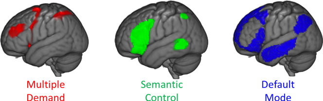
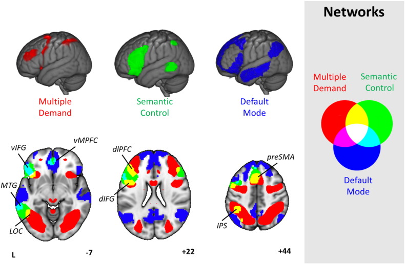
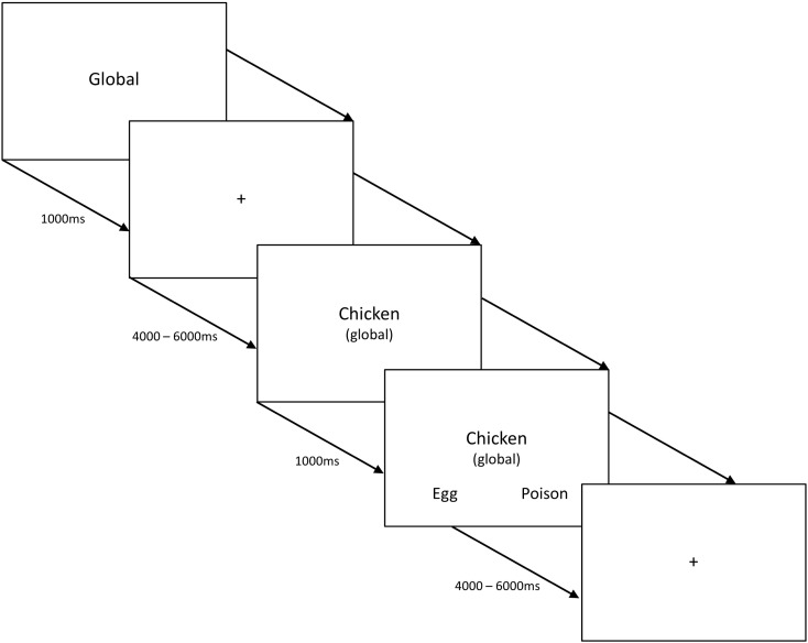
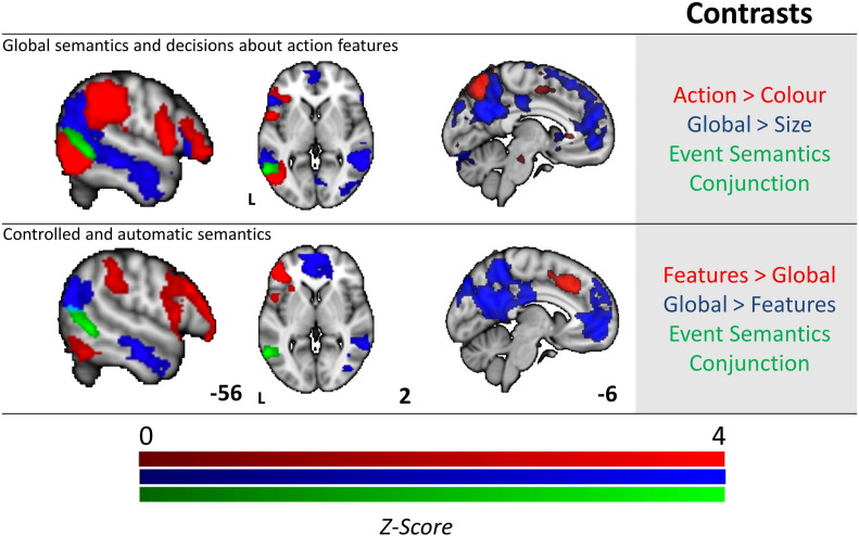
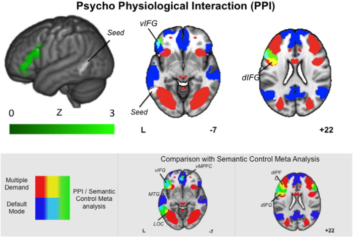
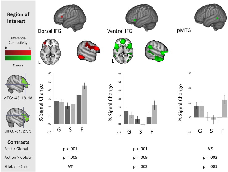
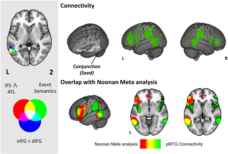
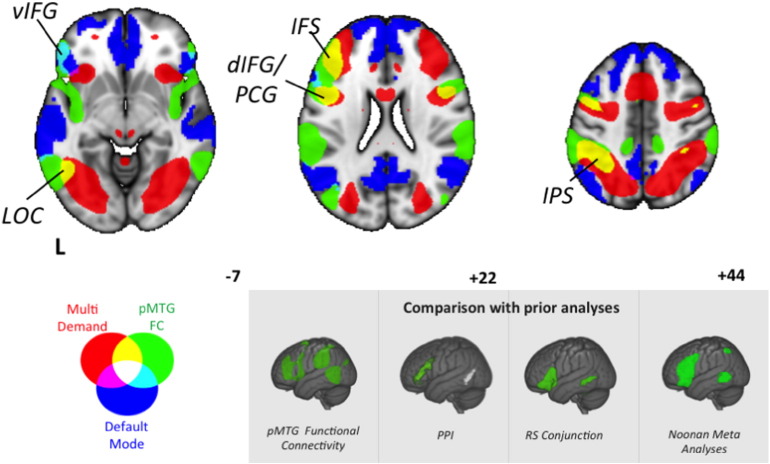
Making sense of the world around us depends upon selectively retrieving information relevant to our current goal or context. However, it is unclear whether selective semantic retrieval relies exclusively on general control mechanisms recruited in demanding non-semantic tasks, or instead on systems specialised for the control of meaning. One hypothesis is that the left posterior middle temporal gyrus (pMTG) is important in the controlled retrieval of semantic (not non-semantic) information; however this view remains controversial since a parallel literature links this site to event and relational semantics. In a functional neuroimaging study, we demonstrated that an area of pMTG implicated in semantic control by a recent meta-analysis was activated in a conjunction of (i) semantic association over size judgements and (ii) action over colour feature matching. Under these circumstances the same region showed functional coupling with the inferior frontal gyrus - another crucial site for semantic control. Structural and functional connectivity analyses demonstrated that this site is at the nexus of networks recruited in automatic semantic processing (the default mode network) and executively demanding tasks (the multiple-demand network). Moreover, in both task and task-free contexts, pMTG exhibited functional properties that were more similar to ventral parts of inferior frontal cortex, implicated in controlled semantic retrieval, than more dorsal inferior frontal sulcus, implicated in domain-general control. Finally, the pMTG region was functionally correlated at rest with other regions implicated in control-demanding semantic tasks, including inferior frontal gyrus and intraparietal sulcus. We suggest that pMTG may play a crucial role within a large-scale network that allows the integration of automatic retrieval in the default mode network with executively-demanding goal-oriented cognition, and that this could support our ability to understand actions and non-dominant semantic associations, allowing semantic retrieval to be 'shaped' to suit a task or context.

Keywords: Default mode network; Memory retrieval; Multidemand network; Posterior middle temporal gyrus; Semantic control.


Figures

Image 1
Graphical abstract
Fig. 1
Fig. 1
Spatial maps of the Default Mode Network (DMN, blue, from Yeo et al., 2011), Multiple-Demand Network (MDN, red, from Fedorenko et al., 2013) and Semantic Control Network (green, from Noonan et al., 2013), presented on a rendered MNI-152 brain and on axial, coronal, and sagittal slices. The key for overlapping areas between different networks is presented on the right hand side of the figure. Images are shown with fully saturated colours to maximise the visibility of the overlapping regions. Regions implicated in semantic control and also found in the MDN include dlPFC (dorsolateral prefrontal cortex), dIFG (dorsal inferior frontal gyrus), pre-SMA (pre-supplementary motor area), IPS (intraparietal sulcus) and LOC (lateral occipital cortex). Regions implicated in semantic control and also found in the DMN include vIFG (ventral inferior frontal gyrus); vMPFC (ventral medial prefrontal cortex) and pMTG (posterior middle temporal gyrus).
Fig. 2
Fig. 2
Example trial structure for all conditions in the task-based fMRI study. The study employed a 2 × 3 design, with three types of judgements (about global semantic associations, size feature matching and specific feature matching) for animal and tool concepts.
Fig. 3
Fig. 3
Functional activation for the conjunction of global semantic decisions and action features (top row), and the contrast between easy and hard semantic decisions (bottom row). The top row displays activation for global semantic associations > size feature selection (blue), and for tool action feature selection > animal colour feature selection (red). The conjunction revealing the shared activation between these contrasts (event/relational semantics) is shown in green. The bottom row shows the contrast of hard > easy semantic decisions (specific feature selection trials > easier global association judgements, in red) and easy > hard semantic decisions (the reverse contrast, in blue). The event semantics conjunction from the top row is overlaid (in green). All contrasts and conjunctions are cluster corrected for multiple comparisons (inclusion threshold z = 2.3, cluster significance = p < .05).
Fig. 4
Fig. 4
The rendered image shows the results of the psychophysiological interaction (PPI) analysis for the region showing common activation for the global semantic decisions and action features (green). The axial slices illustrate regions of overlap between this spatial map and the default mode network (DMN) and the multiple demand network (MDN). For ease of visual comparison the grey box presents the overlap between the meta-analysis of Semantic control and the same pair of large-scale networks. The PPI was cluster corrected for multiple comparisons (inclusion threshold z = 2.3, cluster significance = p < .05).
Fig. 5
Fig. 5
Resting state functional connectivity and structural connectivity (tractography) for functional peaks identified for hard semantic judgements in a key executive region (− 42 28 16, inferior frontal sulcus — IFS, in red) and easy semantic judgements in a region linked to semantic representation (− 48 2 − 38, anterior temporal lobe — ATL, in blue). The conjunction for the two connectivity patterns is displayed in green. All contrasts and conjunctions are cluster corrected to control for multiple comparisons (inclusion threshold z = 2.3, cluster significance = p < .05). The left-hand column shows the seed regions, columns 2 and 3 show resting state connectivity and white matter (WM) fibre tracts identified using diffusion MRI for each seed and their overlap are displayed on the right. We present the spatial maps from the Noonan et al. (2013) meta-analysis and from the prior PPI analysis to facilitate visual comparison of these three networks. Note the colour bar does not refer to the DTI Images which were corrected using randomise and are presented as fully saturated maps.
Fig. 6
Fig. 6
The upper panel shows the difference in resting-state connectivity between the dorsal (red) and ventral (green) inferior frontal gyrus (IFG) peaks from Badre et al. (2005), presented on MNI-152 axial and coronal slices. All difference maps are cluster corrected to control for multiple comparisons (inclusion threshold z = 2.3, cluster significance = p < .05). Colour bars represent the strength of the difference in the connectivity profiles between the two seed regions in LIFG. The lower panel shows the percent signal change for all experimental conditions extracted from 8 mm regions of interest (ROIs) for dorsal and ventral IFG from Badre et al. (2005) and for posterior middle temporal gyrus (pMTG) from Noonan et al. (2013). For ease of visual presentation we present the overlap between the seed regions in IFG and the resting state connectivity presented in Fig. 5 in the grey box. The black bars represent percentage signal change for the animal conditions, and the grey bars represent the signal change for the tool conditions. Error bars correspond to the standard error, with p values for between-condition t-tests presented below. G = global associations. S = size feature matching. F = specific feature matching.
Fig. 7
Fig. 7
The left hand panel shows overlapping clusters in posterior middle temporal gyrus (pMTG) across different contrasts and analyses; (i) the event semantics task-based conjunction (green, reproduced from Fig. 3), (ii) the inferior frontal sulcus (IFS) and anterior temporal lobe (ATL) connectivity conjunction (red, reproduced from Fig. 5), and (iii) the difference between ventral left inferior frontal gyrus (vLIFG) and dorsal LIFG (dLIFG) connectivity (blue, reproduced from Fig. 6). In the main figure, the top row displays the Neurosynth functional connectivity pattern for a seed corresponding to the centre of gravity (COG) for the cluster where all three contrasts overlap. The bottom row compares this pattern for pMTG (in green) with the Noonan et al. (2013) semantic control meta-analysis from Fig. 1 (in red). Regions that fall within both maps are shown in yellow. Images are shown with fully saturated colours to maximise the visibility of the overlapping regions.
Fig. 8
Fig. 8
Functional connectivity of posterior middle temporal gyrus (pMTG; in green, reproduced from Fig. 7) contrasted with the multiple-demand network (MDN; in red) and default mode network (DMN; in blue). It can be seen that the connectivity of the pMTG intersects with the MDN and DMN in similar regions as the Noonan meta-analysis (see subpanel). Images are shown with fully saturated colours to maximise the visibility of the overlapping regions. vIFG = ventral inferior frontal gyrus; LOC = lateral occipital cortex; IFS = inferior frontal sulcus; dIFG/PCG = dorsal inferior frontal gyrus/precentral gyrus; IPS = intraparietal sulcus. For ease of visual comparison in the grey panel we show the spatial maps generated through our prior three analyses, as well the Noonan meta-analysis.

References

    1. Badre D., D'esposito M. Functional magnetic resonance imaging evidence for a hierarchical organization of the prefrontal cortex. J. Cogn. Neurosci. 2007;19:2082–2099. - PubMed
    1. Badre D., Poldrack R.A., Paré-Blagoev E.J., Insler R.Z., Wagner A.D. Dissociable controlled retrieval and generalized selection mechanisms in ventrolateral prefrontal cortex. Neuron. 2005;47:907–918. - PubMed
    1. Baker D.H., Karapanagiotidis T., Coggan D.D., Wailes-Newson K., Smallwood J. Brain networks underlying bistable perception. NeuroImage. 2015;119:229–234. - PubMed
    1. Behrens T.E.J., Woolrich M.W., Jenkinson M., Johansen-Berg H., Nunes R.G., Clare S., Matthews P.M., Brady J.M., Smith S.M. Characterization and propagation of uncertainty in diffusion-weighted MR imaging. Magn. Reson. Med. 2003;50:1077–1088. - PubMed
    1. Behzadi Y., Restom K., Liau J., Liu T.T. A component based noise correction method (CompCor) for BOLD and perfusion based fMRI. NeuroImage. 2007;37:90–101. - PMC - PubMed