. 2016 Jul;90(1):123-34.
doi: 10.1016/j.kint.2016.03.023. Epub 2016 May 25.

Von Willebrand factor regulates complement on endothelial cells

Affiliations

Von Willebrand factor regulates complement on endothelial cells

Damien G Noone et al. Kidney Int. 2016 Jul.

Abstract

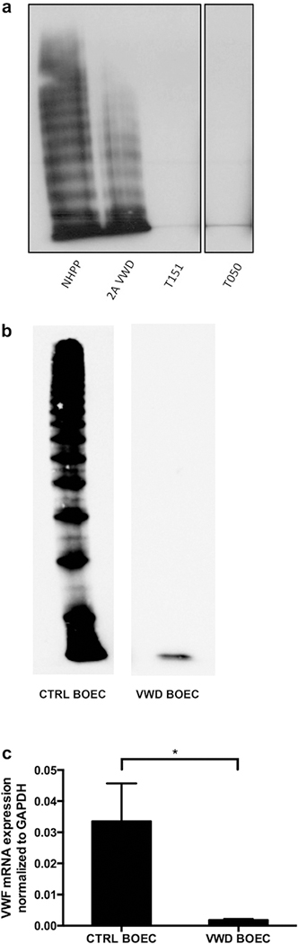
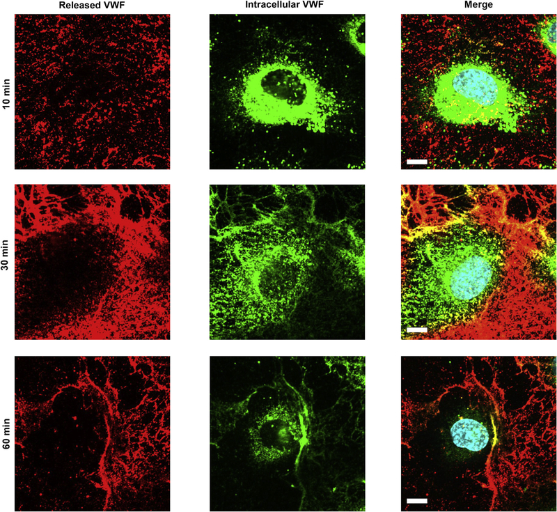
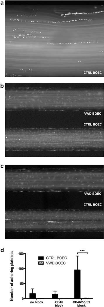
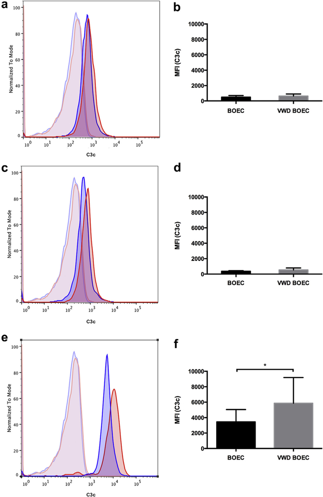
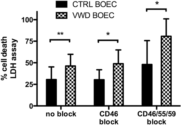
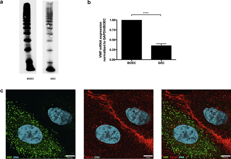
Atypical hemolytic uremic syndrome and thrombotic thrombocytopenic purpura have traditionally been considered separate entities. Defects in the regulation of the complement alternative pathway occur in atypical hemolytic uremic syndrome, and defects in the cleavage of von Willebrand factor (VWF)-multimers arise in thrombotic thrombocytopenic purpura. However, recent studies suggest that both entities are related as defects in the disease-causing pathways overlap or show functional interactions. Here we investigate the possible functional link of VWF-multimers and the complement system on endothelial cells. Blood outgrowth endothelial cells (BOECs) were obtained from 3 healthy individuals and 2 patients with Type 3 von Willebrand disease lacking VWF. Cells were exposed to a standardized complement challenge via the combination of classical and alternative pathway activation and 50% normal human serum resulting in complement fixation to the endothelial surface. Under these conditions we found the expected release of VWF-multimers causing platelet adhesion onto BOECs from healthy individuals. Importantly, in BOECs derived from patients with von Willebrand disease complement C3c deposition and cytotoxicity were more pronounced than on BOECs derived from normal individuals. This is of particular importance as primary glomerular endothelial cells display a heterogeneous expression pattern of VWF with overall reduced VWF abundance. Thus, our results support a mechanistic link between VWF-multimers and the complement system. However, our findings also identify VWF as a new complement regulator on vascular endothelial cells and suggest that VWF has a protective effect on endothelial cells and complement-mediated injury.

Keywords: atypical hemolytic uremic syndrome; blood outgrowth endothelial cells; complement; thrombotic microangiopathy; thrombotic thrombocytopenic purpura; von Willebrand factor.


Conflict of interest statement

DISCLOSURE

All the authors declared no competing interests.

Figures

Figure 1|
Figure 1|. Complement regulators are present on control and VWD BOECs.
By immunofluorescence (a–f) and flow cytometry (gl) the surface expression of complement regulators CD46, CD55, and CD59 was detected. (ac) Control blood outgrowth endothelial cells (BOECs) and (df) von Willebrand disease (VWD) BOECs were seeded on cover slips, stained for CD46 (a,d), CD55 (b,e), and CD59 (c,f) and the representative secondary antibody (Alexa Fluor 488, green), and they were imaged using a fluorescence microscope. Cell nuclei were stained using Hoechst stain (blue). (gl) For flow cytometry, cells were trypsinized off a 6-well plate, and incubated with primary antibody (CD46, CD55, or CD59) and respective secondary Alexa Fluor 488. Surface expression of complement regulators was acquired using an Attune Acoustic Focusing Cytometer (Invitrogen) and analyzed using FlowJo software. A similar surface expression of complement regulators on VWD BOECs (blue) compared to control BOECs (red) was observed, as shown in representative images (gi). The unstained controls are displayed in light blue for VWD BOECs and light red for control BOECs. (jl) Comparison of the median fluorescence intensity (MFI) of 3 experiments (n = 3 for control BOECs and n = 2 for VWD BOECs) did not show a significant difference in surface expression of CD46, CD55, and CD59. (P = 0.7; 2-way analysis of variance.)
Figure 2|
Figure 2|. VWD BOECs lack VWF.
(a) Multimer analysis of the Type 3 von Willebrand disease (VWD) patient plasma (T151 and T050), compared with normal human pooled plasma (NHPP) and Type 2A VWD plasma. Both Type 3 VWD plasma samples lack von Willebrand factor (VWF)-multimers. Images are taken from the same gel with the black lines indicating separation from lanes that were not included in this study. (b) Detection of VWF in control and VWD blood outgrowth endothelial cell (BOECs) showed a decreased amount of protein in lysates of VWD patient BOECs. (c) mRNA expression levels of VWF were determined by RT-qPCR. VWD BOECs showed minimal VWF mRNA expression (*P < 0.05; paired t-test). VWF mRNA levels were normalized to glyceraldehyde-3-phosphate dehydrogenase (GAPDH).
Figure 3|
Figure 3|. Complement fixation results in VWF release.
Von Willebrand factor (VWF) release from control blood outgrowth endothelial cells (BOECs) was detected via immunofluorescence. Control BOECs were treated with CD46, CD55, and CD59 antibody for 20 minutes, followed by 50% normal human serum for 10, 30, and 60 minutes. Cells were fixed with 4% paraformaldehyde, followed by sheep anti-VWF (red). Subsequently, cells were permeabilized with 0.2% Triton in phosphate-buffered saline and incubated with rabbit anti-VWF (green). VWF staining before permeabilization identifies secreted VWF, VWF staining after permeabilization intracellular VWF. Images were taken using an IX81 inverted fluorescence microscope (Olympus Corp., Tokyo, Japan) with a 60/1.35 oil immersion objective and a C9100–13 back-thinned EM-CCD camera (Hamamatsu Photonics, Hamamatsu City, Shizuoka Pref., Japan) with a CSU X1 spinning disk confocal scan head (Yogokawa, Yokogawa Canada, Inc., Alberta, Canada). Bar = 10 μm.
Figure 4|
Figure 4|. Complement-induced platelet adhesion.
(a) In a fluidic system platelet adhesion on control blood outgrowth endothelial cells (BOECs) exposed to complement (treatment as in c) occurred on von Willebrand factor (VWF) strings (original magnification X20). (b,c) Von Willebrand disease (VWD) (upper channel) and control BOECs (lower channel) were exposed to 50% normal human serum for 1 hour without (b) or with (c) CD46, CD55, and CD59 treatment; platelets (15 × 107 per ml, 100 μl per well, in Tyrodes buffer) were perfused at 2 dynes/cm2. No platelet adhesion was observed on VWD BOECs. (d) Platelet adhesion was analyzed by counting adherent platelets of 4 random pictures per channel using ImageJ software. Significantly more platelets were seen to adhere in control BOECs as compared to VWD BOECs devoid of VWF. (N = 3; ***P < 0.001; 2-tailed analysis of variance; Sidak's multiple comparisons test.) Pictures were taken with a Nikon Eclipse Ti camera at X4 (b,c) and X20 (a) original magnification after 5 minutes.
Figure 5|
Figure 5|. Complement deposition on VWD BOECs.
Surface deposition of C3b detected via a polyclonal rabbit anti-C3c antibody was acquired using an Attune Acoustic Focusing Cytometer (Invitrogen) after gating for live and single cells. Experiments were performed using 2 different controls and 2 different von Willebrand disease (VWD) blood outgrowth endothelial cells (BOECs). (a,c,e) Representative figures for control (blue) and VWD BOECs (red) when treated (a) with 50% normal human serum (NHS) alone, (c) after CD46 block, and (e) after CD46, CD55, and CD59 block. Unstained controls for control (light blue) and VWD BOECs (light red). (b,d,f) The median fluorescence intensity (MFI) was calculated using FlowJo software after subtraction of MFI of the unstained sample. No difference was observed when control and VWD BOECs were treated (b) with 50% NHS alone or (d) after CD46 block. (f) A significant increase of C3c deposition was seen in VWD BOECs compared to control BOECs after functional blockade of complement regulators CD46, CD55, CD59, and 50% NHS for 1 hour in both. (N = 5; *P < 0.05; paired t-test.)
Figure 6|
Figure 6|. Complement-mediated cytotoxicity in VWD BOECs.
Cell death was measured by detection of lactate dehydrogenase (LDH) release in supernatant of control and von Willebrand disease (VWD) blood outgrowth endothelial cells (BOECs) after incubation with 10% normal human serum in serum-free media (SFM) for 4 hours of pretreatment with none, only CD46, or a combination of CD46, CD55, and CD59 blocking antibodies. Cell death was calculated using a standard curve and normalized to positive control (100%), obtained adding lysis buffer 45 minutes prior to incubation end. Data were gathered from 3 different experiments (mean of 4–8 wells per plate) using 2 different control BOECs and 2 different VWD BOECs. A more profound increase of cytotoxicity was observed in VWD BOECs compared to control BOECs in all conditions. (*P < 0.05; **P < 0.01; paired t-test.)
Figure 7|
Figure 7|. Glomerular endothelial cells express less VWF.
(a) Control and glomerular endothelial cell (GEC) lysates were resolved by 1.6% LGT agarose gel and probed for von Willebrand factor (VWF). A lower expression of VWF was seen in GECs. (b) mRNA levels of VWF were measured by RT-qPCR in control BOECs and GECs, and VWF mRNA levels of GECs were only 35% of levels found in control BOECs (****P < 0.0001, 2-tailed t-test, N = 3). GAPDH, glyceraldehyde-3-phosphate dehydrogenase. (c) Immunofluorescence labeling of GECs with antibodies detecting VWF (1:1000, green) and VE-cadherin as endothelial cell marker (1:200, red) revealed partial expression of VWF in GECs. Image was taken using an Olympus IX81 inverted fluorescence microscope with a 60/1.35 oil immersion objective and a Hamamatsu C9100–13 back-thinned EM-CCD camera with a Yokogawa CSU X1 spinning disk confocal scan head.

References

    1. Noris M, Remuzzi G. Atypical hemolytic-uremic syndrome. N Engl J Med. 2009;361:1676–1687. - PubMed
    1. Tsai HM. Pathophysiology of thrombotic thrombocytopenic purpura. Int J Hematol. 2010;91:1–19. - PMC - PubMed
    1. George JN. Measuring ADAMTS13 activity in patients with suspected thrombotic thrombocytopenic purpura: when, how, and why? Transfusion. 2015;55:11–13. - PMC - PubMed
    1. Tsai HM. Untying the knot of thrombotic thrombocytopenic purpura and atypical hemolytic uremic syndrome. Am J Med. 2013;126:200–209. - PubMed
    1. Chapin J, Eyler S, Smith R, et al. Complement factor H mutations are present in ADAMTS13-deficient, ticlopidine-associated thrombotic microangiopathies. Blood. 2013;121:4012–4013. - PubMed

Publication types

MeSH terms

LinkOut - more resources