. 2016 Jun 30;166(1):209-21.
doi: 10.1016/j.cell.2016.05.011. Epub 2016 May 26.

Sensory Neurons that Detect Stretch and Nutrients in the Digestive System

Affiliations

Sensory Neurons that Detect Stretch and Nutrients in the Digestive System

Erika K Williams et al. Cell. .

Erratum in

Abstract

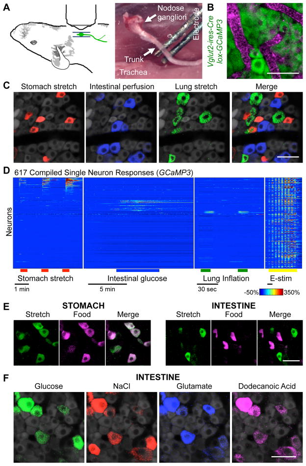
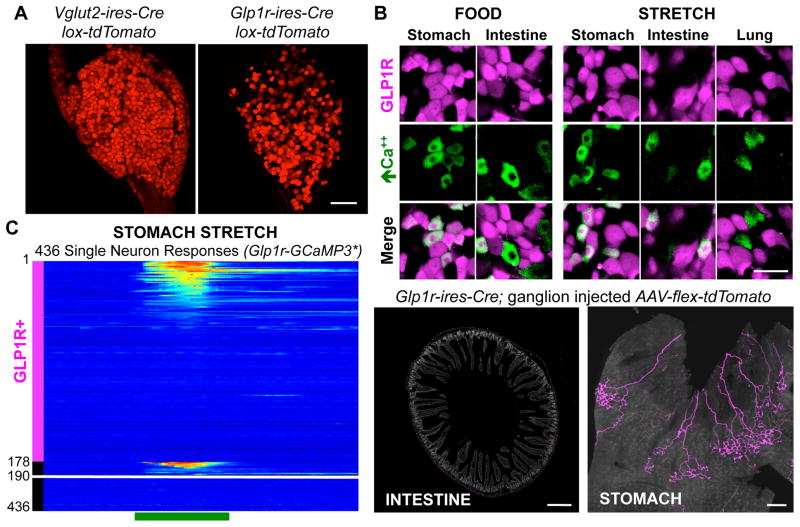
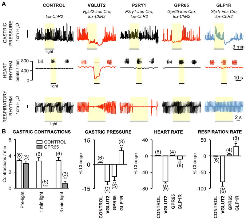
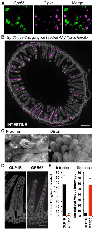
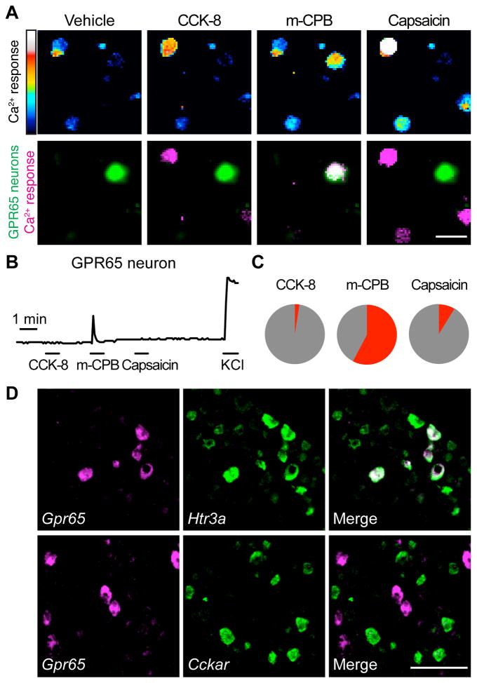
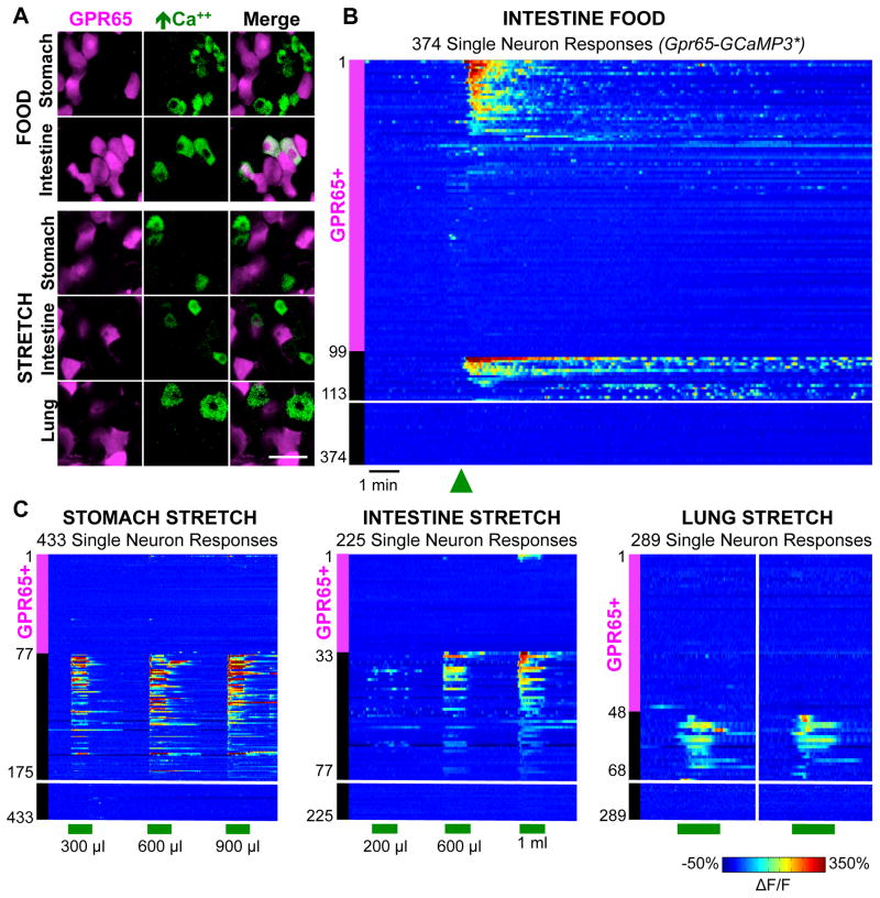
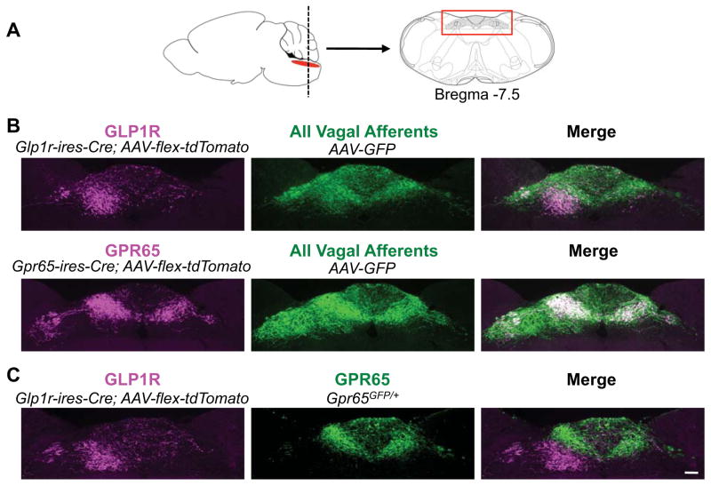
Neural inputs from internal organs are essential for normal autonomic function. The vagus nerve is a key body-brain connection that monitors the digestive, cardiovascular, and respiratory systems. Within the gastrointestinal tract, vagal sensory neurons detect gut hormones and organ distension. Here, we investigate the molecular diversity of vagal sensory neurons and their roles in sensing gastrointestinal inputs. Genetic approaches allowed targeted investigation of gut-to-brain afferents involved in homeostatic responses to ingested nutrients (GPR65 neurons) and mechanical distension of the stomach and intestine (GLP1R neurons). Optogenetics, in vivo ganglion imaging, and genetically guided anatomical mapping provide direct links between neuron identity, peripheral anatomy, central anatomy, conduction velocity, response properties in vitro and in vivo, and physiological function. These studies clarify the roles of vagal afferents in mediating particular gut hormone responses. Moreover, genetic control over gut-to-brain neurons provides a molecular framework for understanding neural control of gastrointestinal physiology.


Figures

Figure 1
Figure 1. In vivo imaging of vagal sensory neurons
(A) Cartoon and photograph of imaging preparation. (B) Wholemount image of GCaMP3 fluorescence (green) and blood vessels (magenta, intravenous Evans Blue) in vagal ganglia. (C) GCaMP3 fluorescence signal in vagal ganglia during stomach stretch (red), intestinal perfusion (blue), and lung stretch (green). (D) Time-resolved responses (ΔF/F, color scale) of 617 neurons (1 neuron per row) to stimuli indicated. GCaMP3 fluorescence signal in vagal ganglia during (E) organ stretch (nitrogen, saline), food injection (liquid diet), or (F) intestinal perfusion of glucose (1 M, saline), sodium chloride (500 mM, saline), sodium glutamate (500 mM, saline) and dodecanoic acid (25 mM, saline and conjugated mouse bile). Scale bars: 50 μm. See also Figures S1, S2, and Movie S1.
Figure 2
Figure 2. Vagal GLP1R neurons are mechanoreceptors
(A) Whole mount tdTomato fluorescence in vagal ganglia from knock-in mice, scale bar: 100 μm. (B) Tomato fluorescence indicating GLP1R neurons (magenta) and GCaMP3 fluorescence responses to stimuli indicated (green) in vagal ganglia of Glp1r-GCaMP3* mice. (C) Time-resolved responses (ΔF/F, color scale) of 178 GLP1R neurons, and 258 other neurons (12 depicted which responded) to gastric distension induced by nitrogen perfusion. (D) AAV mapping of GLP1R neuron projections in intestine and stomach. scale bars: 500 μm (left), 1 mm (right). See also Figures S3, S4.
Figure 3
Figure 3. Optogenetic control of gut motility
(A) Physiological responses to optogenetic activation (yellow bar) of vagal sensory neuron subtypes. (B) Quantifying physiological changes to neuron subtype stimulation (mean ± sem, n=3–8, *p<.05, **p<.01, ***p<.001). See also Figure S5.
Figure 4
Figure 4. GPR65 neurons target intestinal villi
(A) Two-color FISH in vagal ganglia reveals expression of Gpr65 and Glp1r in different sensory neurons, scale bar: 100 μm. (B) Vagal sensory neuron projections were mapped by infecting vagal ganglia of Gpr65-ires-Cre mice with AAV-flex-tdTomato. Terminals were visualized by immunofluorescence of duodenum (cryosections) and intestinal architecture visualized with DAPI (grey), scale bar: 500 μm. (C) Wholemount fluorescence of nerve terminals in an en face preparation of proximal (< 1 cm from pylorus) and distal (4 cm from pylorus) intestinal villi after injecting vagal ganglia of Vglut2-ires-Cre mice with AAV-flex-tdTomato. (D) High magnification image of villi innervation, scale bar: 100 μm. (E) Numbers of intestinal villi and gastric enteric ganglia innervated by vagal sensory neuron types were counted, and for villi, normalized using a Cre-independent reporter (mean ± sem, n=6, **p<.01). See also Figure S5.
Figure 5
Figure 5. GPR65 neurons respond in vitro to an HTR3A agonist
(A) Calcium responses by Fura-2 imaging of dissociated vagal sensory neurons from Gpr65GFP/+ mice to CCK-8 (10 nM), the HTR3A agonist m-chlorophenylbiguanide (mCPB, 100 μM) and capsaicin (1 μM), scale bar: 40 μm. Top: Fura-2 excitation; bottom: GFP fluorescence (green) and calcium responses (magenta). (B) Representative responses of a GPR65 neuron. (C) Pie chart indicating percentage of GPR65 neurons activated (red) by each ligand. (D) Two color FISH in vagal ganglia. Scale bar: 100 μm. See also Figure S6.
Figure 6
Figure 6. GPR65 neurons detect intestinal nutrients in vivo
(A) In vivo imaging of vagal ganglia in Gpr65-GCaMP3* mice showing GCaMP3 responses (green) of GPR65 neurons (magenta) to stimuli indicated. (B) Rows indicate time-resolved responses (ΔF/F, color coded) of single neurons in Gpr65-GCaMP3* mice to stimuli (green bars: 15 seconds). Magenta and black bars represent tdTomato-positive and negative neurons. Only some unresponsive tdTomato-negative neurons are depicted; numbers at Y-axis base indicate total number of viable imaged neurons. See also Figure S6.
Figure 7
Figure 7. Visualizing brainstem innervation
(A) Vagal sensory neuron axons were analyzed in a brainstem region (red box) containing the NTS and area postrema. (B) Vagal ganglia of Glp1r-ires-Cre and Gpr65-ires-Cre mice were infected with AAV-flex-tdTomato and AAV-GFP for immunofluorescence-based detection of Cre-containing (magenta) and all (green) vagal sensory neuron axon types. (C) Vagal ganglia of Glp1r-ires-Cre; Gpr65GFP/+ mice were infected with AAV-flex-tdTomato for simultaneous visualization of GLP1R (magenta) and GPR65 (green) axons, scale bar: 100 μm. See also Figure S7.

Comment in

References

    1. Abbott CR, Monteiro M, Small CJ, Sajedi A, Smith KL, Parkinson JR, Ghatei MA, Bloom SR. The inhibitory effects of peripheral administration of peptide YY(3-36) and glucagon-like peptide-1 on food intake are attenuated by ablation of the vagal-brainstem-hypothalamic pathway. Brain research. 2005;1044:127–131. - PubMed
    1. Aponte Y, Atasoy D, Sternson SM. AGRP neurons are sufficient to orchestrate feeding behavior rapidly and without training. Nature neuroscience. 2011;14:351–355. - PMC - PubMed
    1. Baggio LL, Drucker DJ. Biology of incretins: GLP-1 and GIP. Gastroenterology. 2007;132:2131–2157. - PubMed
    1. Bai L, Lehnert BP, Liu J, Neubarth NL, Dickendesher TL, Nwe PH, Cassidy C, Woodbury CJ, Ginty DD. Genetic Identification of an Expansive Mechanoreceptor Sensitive to Skin Stroking. Cell. 2015;163:1783–1795. - PMC - PubMed
    1. Barretto RP, Gillis-Smith S, Chandrashekar J, Yarmolinsky DA, Schnitzer MJ, Ryba NJ, Zuker CS. The neural representation of taste quality at the periphery. Nature. 2015;517:373–376. - PMC - PubMed

Publication types