. 2017 May;14(5):465-475.
doi: 10.1038/cmi.2016.24. Epub 2016 May 30.

Exosomes mediate hepatitis B virus (HBV) transmission and NK-cell dysfunction

Affiliations

Exosomes mediate hepatitis B virus (HBV) transmission and NK-cell dysfunction

Yinli Yang et al. Cell Mol Immunol. 2017 May.

Abstract

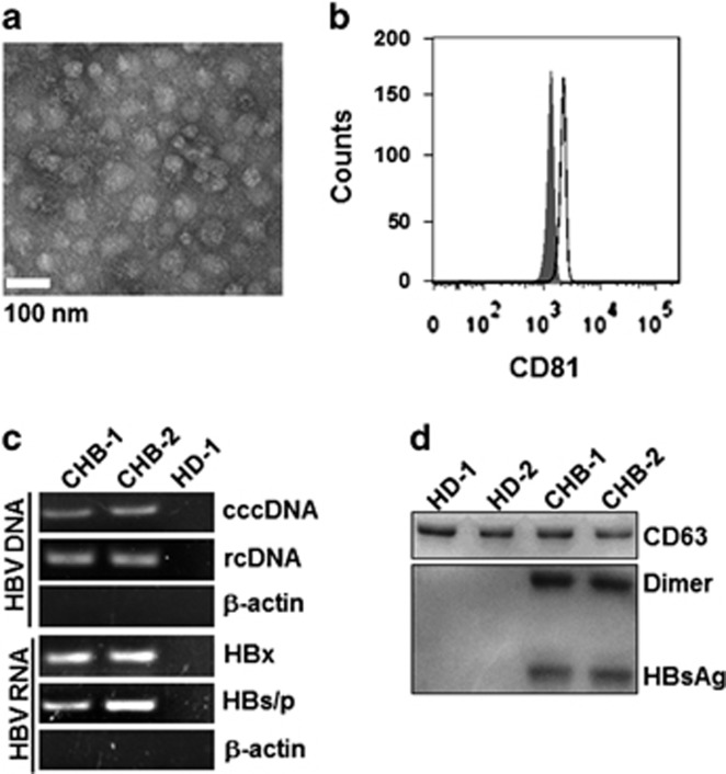
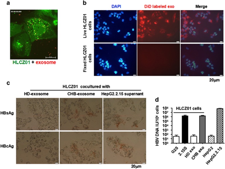
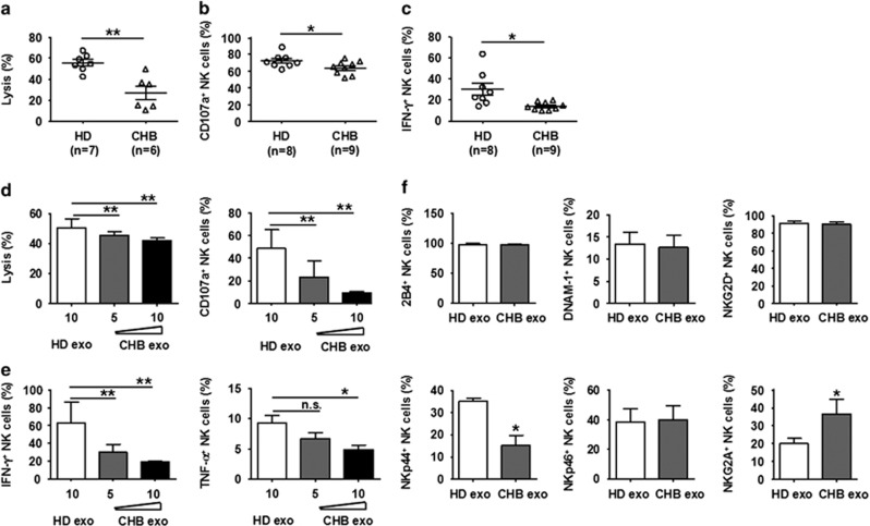
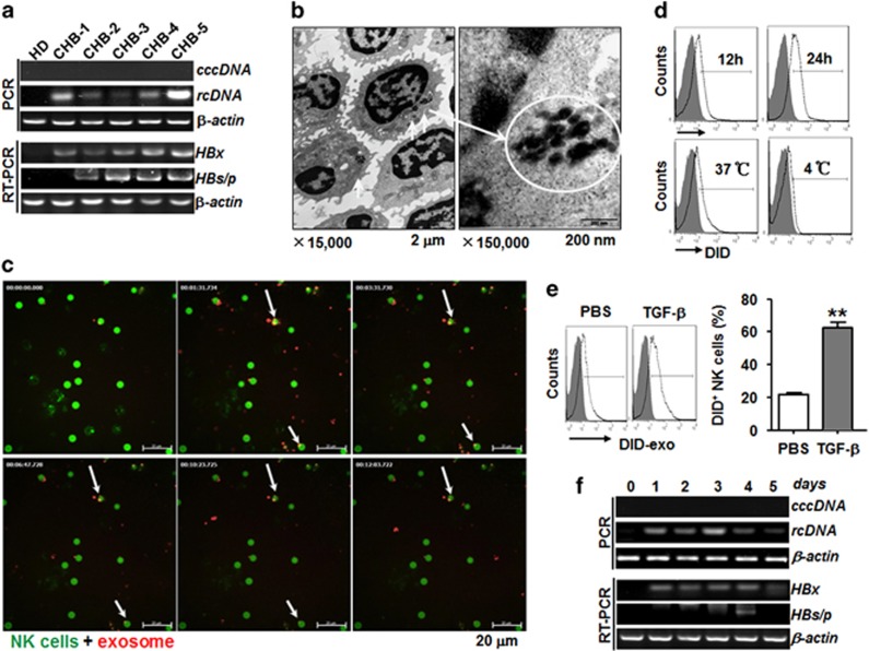
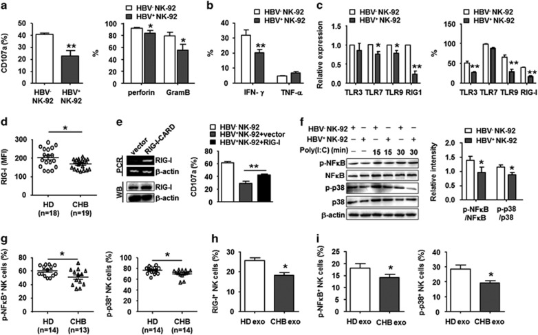
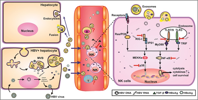
Evidence suggests that exosomes can transfer genetic material between cells. However, their roles in hepatitis B virus (HBV) infection remain unclear. Here, we report that exosomes present in the sera of chronic hepatitis B (CHB) patients contained both HBV nucleic acids and HBV proteins, and transferred HBV to hepatocytes in an active manner. Notably, HBV nucleic acids were detected in natural killer (NK) cells from both CHB patients and healthy donors after exposure to HBV-positive exosomes. Through real-time fluorescence microscopy and flow cytometry, 1,1'-dioctadecyl-3,3,3',3',-tetramethylindodicarbocyanine, 4-chlorobenzenesulfnate salt (DiD)-labeled exosomes were observed to interact with NK cells and to be taken up by NK cells, which was enhanced by transforming growth factor-β treatment. Furthermore, HBV-positive exosomes impaired NK-cell functions, including interferon (IFN)-γ production, cytolytic activity, NK-cell proliferation and survival, as well as the responsiveness of the cells to poly (I:C) stimulation. HBV infection suppressed the expression of pattern-recognition receptors, especially retinoic acid inducible gene I (RIG-I), on NK cells, resulting in the dampening of the nuclear factor κB(NF-κB) and p38 mitogen-activated protein kinase pathways. Our results highlight a previously unappreciated role of exosomes in HBV transmission and NK-cell dysfunction during CHB infection.


Conflict of interest statement

The authors declare no conflict of interest.

Figures

Figure 1
Figure 1
Exosomes derived from CHB patients contained viral components. (a) Electron microscope image of purified exosomes from CHB serum. Scale bar, 100 nm. (b) CD81 protein was used to identify the exosomes isolated from CHB serum using flow cytometry. The shaded histograms represent the isotype control. (c) HBV DNA (rcDNA and cccDNA) and RNA (HBx and HBs/p) in exosomes isolated from CHB serum were detected using PCR and RT-PCR, respectively. (d) Western blot analysis of HBsAg and CD63 in exosomes from HD and CHB patient serum (10 μg/well). (Because of the limited volume of each sample, six fresh serum samples from CHB patients or healthy donors were harvested and mixed for exosome isolation). One representative of at least three independent experiments is shown. CHB, chronic hepatitis B; HBV, hepatitis B virus; DNA, deoxyribonucleic acid; rcDNA, relaxed circular DNA; cccDNA, closed-circular DNA; RNA, ribonucleic acid; PCR, polymerase chain reaction; RT-PCR, reverse transcription polymerase chain reaction; HD, healthy donors.
Figure 2
Figure 2
Exosomes can transmit HBV to uninfected hepatoma cells. (a) The confocal image shows the co-localization of CFSE-labeled HLCZ01 cells (green) incubated with DiD-labeled exosomes (red) for 2 h. Scale bar, 20 μm. (b) Comparison of the ability to take up DiD-labeled exosomes (red) between live HLCZ01 cells and fixed HLCZ01 cells. Scale bar, 20 μm. (c) Immunocytochemical staining of HBsAg and HBcAg proteins in HLCZ01 cells infected with exosomes or HepG2.2.15 supernatant. Scale bar, 20 μm. (d) Quantitative PCR analysis of HBV DNA in HLCZ01 cells treated with HD/CHB exosomes, the supernatant of HepG2 cells or the supernatant of HepG2.2.15 cells at an multiplicity of infection of 50 per cell (n=3). HepG2 and HepG2.2.15 cells were used as negative and positive controls, respectively. The results are representative of at least three independent experiments. The data are expressed as the mean±s.e.m. *P<0.05, **P<0.01. CHB, chronic hepatitis B; DNA, deoxyribonucleic acid; GS, HepG2 supernatant; HBV, hepatitis B virus; HD, healthy donors; 2.15S, HepG2.2.15 supernatant; PCR, polymerase chain reaction.
Figure 3
Figure 3
Exosomes derived from CHB patients affected NK cell function. PBMCs from HD and CHB patients were used to determine NK cell-mediated cytotoxicity against K562 cells using the 3-(4,5-cimethylthiazol-2-yl)-2,5-diphenyl tetrazolium bromide (MTT) method (a), and the production of CD107a (b) and IFN-γ (c) in CD56+CD3 NK cells was analyzed using flow cytometry. (d) NK cells isolated from HD were treated with exosomes (5 or 10 μg/ml) from CHB or HD. Then, the cytotoxicity against K562 cells was determined by CFSE/7AAD assay (left) and CD107a production was tested by flow cytometry (right). (e) IFN-γ and TNF-α production by NK cells treated as in d were determined by flow cytometry. (f) The expression of activating receptors and inhibitory receptors on NK cells treated with CHB exosomes (10 μg/ml) was determined by fluorescence activated cell sorting (FACS) analysis. The results are representative of three independent experiments. The data are expressed as the mean±s.e.m. *P<0.05, **P<0.01. CHB, chronic hepatitis B; HD, healthy donors; NK, natural killer; PBMCs, peripheral blood mononuclear cells.
Figure 4
Figure 4
HBV can be transmitted into NK cells through exosomes. (a) Identification of HBV DNA (rcDNA and cccDNA) and HBV RNA (HBx and HBs/p) in NK cells isolated from HD and CHB patients. (b) Transmission electron microscopy of ultrathin sections was used to observe freshly isolated NK cells from CHB patients with high virus loading. Cytoplasmic inclusion bodies with irregular electron density are shown on the left and middle panels (arrows and loop). Scale bar, 2 μm and 200 nm. (c) Confocal images of the co-localization of CFSE-labeled (green) primary NK cells from HD incubated with DiD-labeled exosomes (red) for 3 h. Scale bar, 20 μm. (d) The uptake of DiD-labeled exosomes by primary NK cells after 12 or 24 h incubation at 37 or 4 °C was determined by flow cytometry. (e) TGF-β (5 ng/ml) enhanced the ability of primary NK cells to take up DiD-labeled exosomes (red). (f) Analysis of HBV DNA (rcDNA and cccDNA) and RNA (HBx and HBs/p) in primary NK cells from HD infected with exosomes derived from CHB serum for 0–5 days. The results are representative of at least three independent experiments. The data are expressed as the mean±s.e.m. *P<0.05, **P<0.01. cccDNA, closed-circular DNA; CHB, chronic hepatitis B; DNA, deoxyribonucleic acid; HBV, hepatitis B virus; HD, healthy donors; NK, natutal killer; rcDNA, relaxed circular DNA; RNA, ribonucleic acid.
Figure 5
Figure 5
HBV components inhibited NK cell function and cell viability. Cytotoxicity was evaluated using a CFSE/7AAD assay (a), and the levels of the degranulation molecule CD107a and the intracellular cytotoxic mediators perforin and GramB (b) in HBV+ NK-92 and HBV NK-92 cells were analyzed using flow cytometry. (c) The intracellular levels of IFN-γ, TNF-α in HBV+ NK-92 and HBV NK-92 cells were analyzed using flow cytometry. (d) The proliferation of HBV+ NK-92 and HBV NK-92 cells was analyzed using an MTT assay. (e) The cell cycle of HBV+ NK-92 and HBV NK-92 cells was analyzed using flow cytometry. (f) The apoptotic rate of HBV+ NK-92 and HBV NK-92 cells after serum starvation was analyzed via flow cytometry using PI and APC-labeled Annexin V. One representative of three independent experiments is shown. The data are expressed as the mean±s.e.m. from at least three independent experiments. *P<0.05, **P<0.01. HBV, hepatitis B virus; NK, natural killer; Star, serum starvation; Untr, untreated.
Figure 6
Figure 6
HBV dampened the expression of RIG-I and its downstream signaling pathway in NK cells. (a) The levels of CD107a and molecules associated with cytolysis, perforin and GramB, in HBV+ NK-92 cells in response to poly (I:C) stimulation were analyzed using flow cytometry. (b) The intracellular levels of IFN-γ and TNF-α in HBV+ NK-92 cells in response to poly (I:C) stimulation were analyzed using flow cytometry. (c) TLR and RIG-I mRNA (left) and protein (right) levels in HBV+ NK-92 and control cells. (d) The RIG-I expression levels in CD56+CD3 NK cells from HD and CHB patients were assessed through flow cytometry. (e) The pcDNA5-RIG-I CARD plasmid was used to transfect HBV+ NK-92 cells (left), and the analysis of the CD107a expression is shown (right). (f) WB blot analysis for NF-κB (p65) and p38 activation in HBV+ NK-92 cells in response to poly (I:C) stimulation (left). The densitometric analysis for p-NF-κB and p-p38 expression is normalized to their total protein levels in NK-92 cells stimulated with poly (I:C) for 15 min, and the control group of unstimulated HBV NK-92 cells was set as 1 (right). (g) NF-κB (p65) and p38 phosphorylation levels in primary CD56+CD3 NK cells from HD and CHB patients. The RIG-I expression (h), NFκB and p38 phosphorylation levels (i) in NK cells from HD were analyzed using flow cytometry after CHB exosome treatment. One representative of at least three independent experiments is shown. The data are expressed as the mean±s.e.m. *P<0.05, **P<0.01. CHB, chronic hepatitis B; HBV, hepatitis B virus; HD, healthy donors; NK, natural killer; WB, western blotting.
Figure 7
Figure 7
Schematic of exosome-mediated HBV transmission and NK cell dysfunction. In CHBV infection, exosomes are derived from the internal vesicles of multivesicular bodies (MVBs) of HBV-infected hepatocytes. MVBs fuse with the plasma membrane and release exosomes that contain HBV components. Then, these exosomes transmit HBV into naive hepatocytes and even NK cells. In NK cells, HBV nucleic acids transmitted by exosomes affect NK cytolytic activity, cytokine production, and cell proliferation. The underlying mechanisms are associated with RIG-I expression and NF-κB and p38 activation.

Comment in

Similar articles

Cited by

References

    1. Lavanchy D. Hepatitis B virus epidemiology, disease burden, treatment, and current and emerging prevention and control measures. J Viral Hepat 2004; 11: 97–107. - PubMed
    1. McMahon BJ. The natural history of chronic hepatitis B virus infection. Hepatology 2009; 49: S45–S55. - PubMed
    1. McMahon BJ. Chronic hepatitis B virus infection. Med Clin North Am 2014; 98: 39–54. - PubMed
    1. Glebe D, Urban S. Viral and cellular determinants involved in hepadnaviral entry. World J Gastroenterol 2007; 13: 22–38. - PMC - PubMed
    1. Yan H, Zhong G, Xu G, He W, Jing Z, Gao Z et al. Sodium taurocholate cotransporting polypeptide is a functional receptor for human hepatitis B and D virus. Elife 2012; 1: e00049. - PMC - PubMed

MeSH terms