. 2016 Feb 13:9721:972106.
doi: 10.1117/12.2207908. Epub 2016 Mar 7.

Extremely sensitive dual imaging system in solid phantoms

Affiliations

Extremely sensitive dual imaging system in solid phantoms

Eran A Barnoy et al. Proc SPIE Int Soc Opt Eng. .

Abstract

Herein we describe promising results from the combination of fluorescent lifetime imaging microscopy (FLIM) and diffusion reflection (DR) medical imaging techniques. Three different geometries of gold nanoparticles (GNPs) were prepared: spheres of 20nm diameter, rods (GNRs) of aspect ratio (AR) 2.5, and GNRs of AR 3.3. Each GNP geometry was then conjugated using PEG linkers estimated to be 10nm in length to each of 3 different fluorescent dyes: Fluorescein, Rhodamine B, and Sulforhodamine B. DR provided deep-volume measurements (up to 1cm) from within solid, tissue-imitating phantoms, indicating GNR presence corresponding to the light used by recording light scattered from the GNPs with increasing distance to a photodetector. FLIM imaged solutions as well as phantom surfaces, recording both the fluorescence lifetimes as well as the fluorescence intensities. Fluorescence quenching was observed for Fluorescein, while metal-enhanced fluorescence (MEF) was observed in Rhodamine B and Sulforhodamine B - the dyes with an absorption peak at a slightly longer wavelength than the GNP plasmon resonance peak. Our system is highly sensitive due to the increased intensity provided by MEF, and also because of the inherent sensitivity of both FLIM and DR. Together, these two modalities and MEF can provide a lot of meaningful information for molecular and functional imaging of biological samples.

Keywords: Gold nanoparticles; biomolecular imaging; diffusion reflection; fluorescence lifetime imaging; gold nanorods; metal enhanced fluorescence; noninvasive detection; tissue-imitating phantoms.


Figures

Figure 1
Figure 1
Scheme depicting the manufacturing process to creating the imaging probe, for both gold nanospheres (GNSs) and gold nanorods (GNRs). Also shown are the chemical structures of the 3 fluorophores used.
Figure 2
Figure 2
Normalized absorption spectra are shown for the range 400–900 nm, for (A) the 3 GNP geometries, (B) Fluorescein compared to the GNPs, (C) Rhodamine B compared to the GNPs, and (D) Sulforhodamine B compared to the GNPs. TEM images of (E) GNRs with peak at 760nm, (F) GNRs with peak at 690nm, and (G) GNSs.
Figure 3
Figure 3
FI enhancement and quenching due to GNP conjugation in solution. FI is depicted in photon counts per millisecond, or kilohertz. (a) Count rate histograms of GNS, GNR690, and GNR760 conjugated to RhB. FI of RhB solution without GNPs is also included in the panel for comparison. (b) Count rate histograms of GNS, GNR690, and GNR760 conjugated to SRB. FI of SRB is included in the panel for comparison. (c) Count rate histograms of GNS, GNR690, and GNR760 conjugated to Flu. FI of Flu is included in the panel for comparison. All solutions contained a fluorophore concentration of 1 μM. All measurements for a given fluorophore are obtained under identical conditions, set-up, and excitation power. Reprinted from: Nano Research, “An Ultra-Sensitive Dual-Mode Imaging System Using Metal-Enhanced Fluorescence in Solid Phantoms”, Vol. 8(12), 2015, p. 3917, Barnoy EA, Fixler D, Popovtzer R, Nayhoz T, Ray K, with permission of Springer.
Figure 4
Figure 4
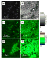
FLIM images of RhB-based phantoms. Depicting only FI (shown in counts of fluorescence events), the images show phantoms containing (a) RhB only and (b) GNS-RhB. Combining FI (shown as brightness) and FLT (shown as color), the images show phantoms containing (c) RhB only, (d) GNS-RhB, (e) GNR690- RhB, and (f) GNR760-RhB. For all images, the gray (brightness) scale bar represents FI in counts per millisecond. The color scale bar displays the FLT range in ns. All images are obtained under identical conditions, set-up, and excitation power. Reprinted from: Nano Research, “An Ultra-Sensitive Dual-Mode Imaging System Using Metal-Enhanced Fluorescence in Solid Phantoms”, Vol. 8(12), 2015, p. 3918, Barnoy EA, Fixler D, Popovtzer R, Nayhoz T, Ray K, with permission of Springer.
Figure 5
Figure 5
Diffusion reflection (DR) results using a 780nm light source, with phantoms measured containing water only (“Base”), and each of the 3 GNP geometries considered. The “Slope” refers to the linear relationship between the intensity of scattered light and the separation distance between light source and detector in the DR system. Each phantom was measured twice, as indicated by the 2 columns.

Similar articles

References

    1. Barnoy EA, Fixler D, Popovtzer R, Nayhoz T, Ray K. An Ultra-Sensitive Dual-Mode Imaging System Using Metal-Enhanced Fluorescence in Solid Phantoms. Nano Res. 2015;8(12):3912–3921. - PMC - PubMed
    1. Eghtedari M, Liopo AV, Copland JA, Oraevsky AA, Motamedi M. Engineering of Hetero-Functional Gold Nanorods for the in Vivo Molecular Targeting of Breast Cancer Cells. Nano Lett. 2009;9:287–291. - PMC - PubMed
    1. von Maltzahn G, Park J, Agrawal A, Bandaru NK, Das SK, Sailor MJ, Bhatia SN. Computationally Guided Photothermal Tumor Therapy Using Long-Circulating Gold Nanorod Antennas. Cancer Res. 2009;69(9):3892–3900. - PMC - PubMed
    1. El-Sayed MA. Some Interesting Properties of Metals Confined in Time and Nanometer Space of Different Shapes. Acc Chem Res. 2001;34(4):257–264. - PubMed
    1. Jain PK, Lee KS, El-Sayed IH, El-Sayed MA. Calculated Absorption and Scattering Properties of Gold Nanoparticles of Different Size, Shape, and Composition: Applications in Biological Imaging and Biomedicine. J Phys Chem B. 2006;110(14):7238–7248. - PubMed

LinkOut - more resources