. 2016:2016:7067691.
doi: 10.1155/2016/7067691. Epub 2016 Apr 27.

Du-Huo-Ji-Sheng-Tang Attenuates Inflammation of TNF-Tg Mice Related to Promoting Lymphatic Drainage Function

Affiliations

Du-Huo-Ji-Sheng-Tang Attenuates Inflammation of TNF-Tg Mice Related to Promoting Lymphatic Drainage Function

Yan Chen et al. Evid Based Complement Alternat Med. 2016.

Abstract

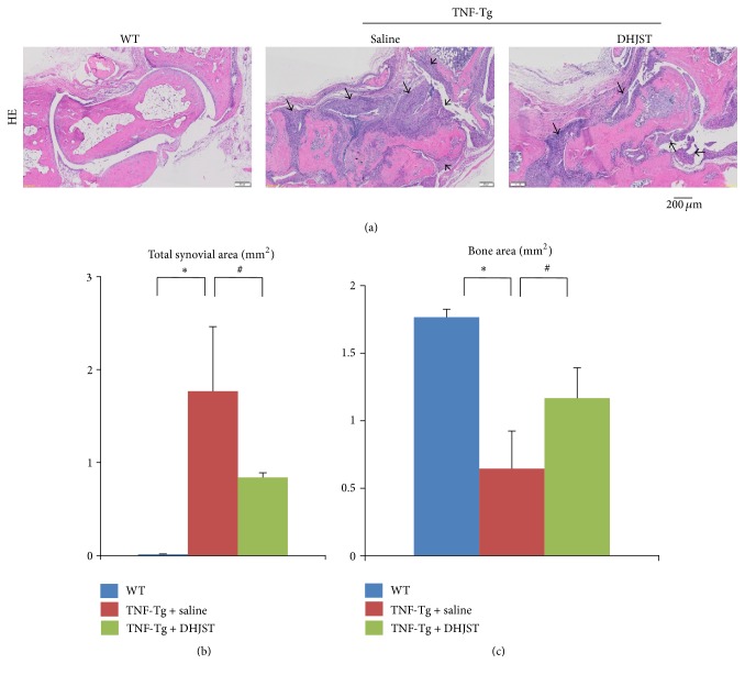
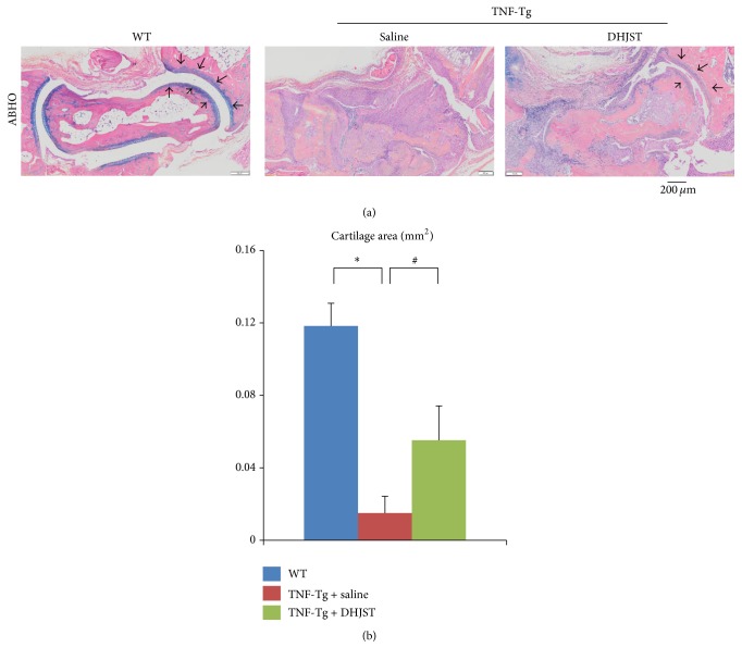
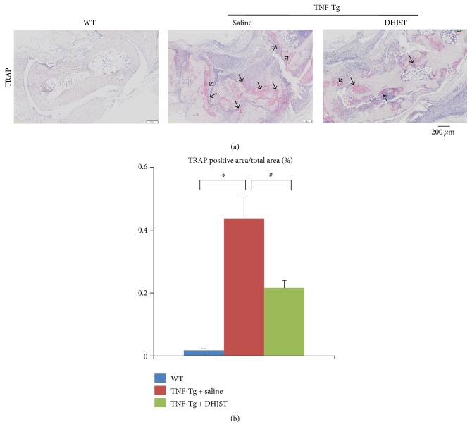
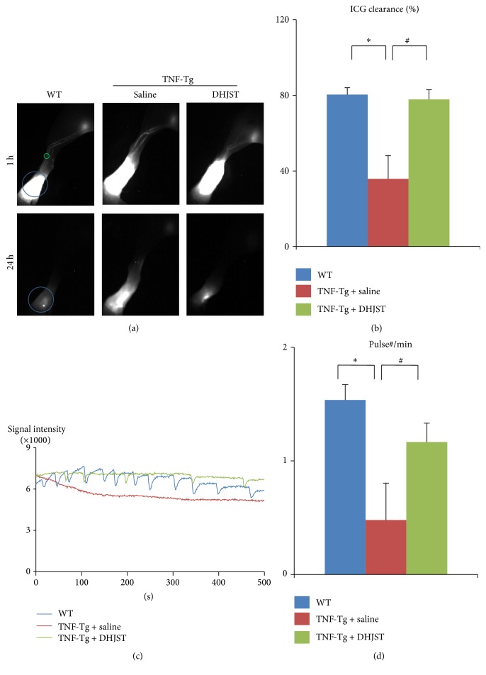
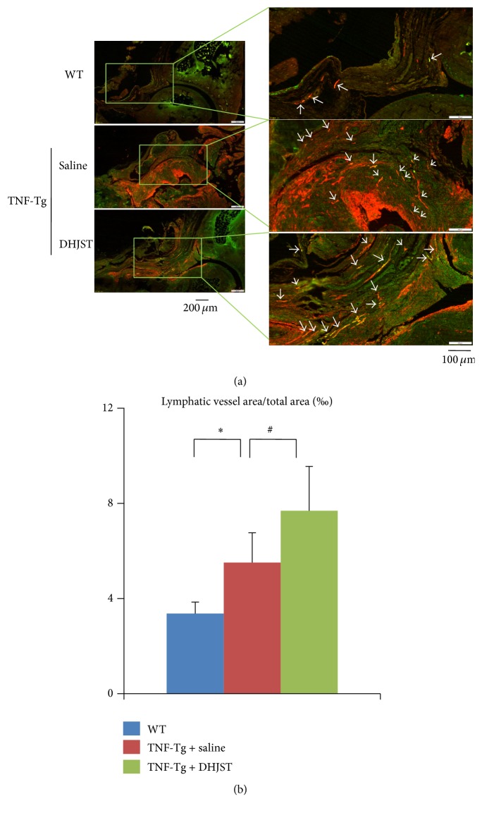
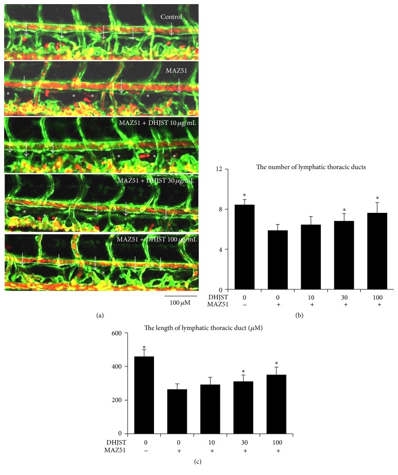
To investigate whether Du-Huo-Ji-Sheng-Tang (DHJST) attenuate inflammation of RA related to lymphatic drainage function in vivo, we treated eight 3-month-old TNF-Tg mice with DHJST (12 g/kg) or the same volume of physiological saline once every day for 12 weeks, and 3-month-old WT littermates were used as negative control. After twelve weeks, we performed NIR-ICG imaging and found that DHJST increased the ICG clearance at the footpad and the pulse of efferent lymphatic vessel between popliteal lymph node and footpad. Histology staining at ankle joints showed that DHJST decreases synovial inflammation, bone erosion, cartilage erosion, and TRAP+ osteoclast area in TNF-Tg mice. Immunohistochemical staining by using anti-Lyve-1 and anti-podoplanin antibody showed that DHJST stimulated lymphangiogenesis in ankle joints of TNF-Tg mice. And zebrafish study suggested that DHJST promoted the formation of lymphatic thoracic duct. In conclusion, DHJST inhibits inflammation severity and promotes lymphangiogenesis and lymphatic drainage function of TNF-Tg mice.


Figures

Figure 1
Figure 1
Du-Huo-Ji-Sheng-Tang reduces inflammation and bone erosion in TNF-Tg mice. Three-month-old TNF-Tg mice were treated with Du-Huo-Ji-Sheng-Tang (12 g/kg/gavage, daily ×12 weeks) or saline. Ankle joints were harvested and subjected to histologic analysis. WT mice were included as control. (a) Representative HE-stained sections show decreased inflammation and bone loss in the Du-Huo-Ji-Sheng-Tang-treated joints. Bar, 200 μm, black arrow indicates inflammatory synovial tissue. Quantitation of bone area (b) and inflammatory synovial area (c). Values are the mean ± SD of 6–8 legs per group. p < 0.05 versus WT; # p < 0.05 versus TNF-Tg + saline.
Figure 2
Figure 2
Du-Huo-Ji-Sheng-Tang reduces cartilage erosion in TNF-Tg mice. (a) Representative ABHO-stained sections show loss of cartilage at ankle joint of saline treated TNF-Tg mice and reduced cartilage erosion in the Du-Huo-Ji-Sheng-Tang-treated joints. Bar, 200 μm, Alcian blue positive staining (black arrow) indicates cartilage at ankle joint. Quantitation of cartilage area (b). Values are the mean ± SD of 6–8 legs per group. p < 0.05 versus WT; # p < 0.05 versus TNF-Tg + saline.
Figure 3
Figure 3
Du-Huo-Ji-Sheng-Tang reduces TRAP+ osteoclast in TNF-Tg mice. (a) Representative TRAP-stained sections show decreased TRAP+ osteoclast number in the Du-Huo-Ji-Sheng-Tang-treated joints. Bar, 200 μm, arrow indicates TRAP+ osteoclast. Quantitation of TRAP+ osteoclast area (b). Values are the mean ± SD of 6–8 legs per group. p < 0.05 versus WT; # p < 0.05 versus TNF-Tg + saline.
Figure 4
Figure 4
Impaired lymphatic function of TNF-Tg mice was rescued by Du-Huo-Ji-Sheng-Tang. Three-month-old TNF-Tg mice were treated with Du-Huo-Ji-Sheng-Tang or saline for 12 weeks and were subjected to ICG-NIR imagining. (a) Representative ICG images show that Du-Huo-Ji-Sheng-Tang increased ICG removal from ankle area. (b) Quantitation of % ICG clearance. Values are mean ± SD of 8–18 legs. (c) Lymphatic pulses were measured at a region of interest. Histogram shows that Du-Huo-Ji-Sheng-Tang restored lymphatic pulses in TNF-Tg mice. (d) Quantitation of lymphatic pulses/min. Values are mean ± SD of 7–18 legs from 4–9 mice. p < 0.05 versus WT mice, # p < 0.05 versus saline treated TNF-Tg group.
Figure 5
Figure 5
DHJST increases lymphangiogenesis in ankle joints of TNF-Tg mice. (a) Three-month DHJST or saline treatment, ankle joints were harvested and subjected to double immunofluorescence staining with anti-LYVE-1 and anti-podoplanin antibodies. Representative LYVE-1 (green) and podoplanin (red) stained ankle sections show that LYVE-1+/podoplanin+ lymphatic vessels (white arrows) are present at synovium and soft tissue surrounding the ankle joints of WT and TNF-Tg mice. (b) Quantitation of LYVE-1+/podoplanin+ lymphatic vessel area inside the areas of ankle joint. Values are the means ± SD of 5-6 legs per group. p < 0.05 versus WT group, # p < 0.05 versus saline treated TNF-Tg group.
Figure 6
Figure 6
Impaired lymphatic thoracic duct formation induced by VEGFR-3 kinase inhibitor (MAZ51) was rescued by Du-Huo-Ji-Sheng-Tang in dose dependent manner. Two dpf zebrafish (fli1:egfp; gata1:DsRed) were treated with 30 μM MAZ51 for 6 hours and then changed to be treated with different doses of Du-Huo-Ji-Sheng-Tang (10–100 μg/mL) for 48 hours. Embryos treated with 0.2% DMSO served as a vehicle control. (a) Representative confocal images show that Du-Huo-Ji-Sheng-Tang increased lymphatic thoracic duct formation of zeberafish, white arrow indicates lymphatic thoracic duct, and white star indicates lack of lymphatic vessel. (b) Quantitation of the number of lymphatic thoracic ducts. (c) Quantitation of the length of lymphatic thoracic duct. Values are mean ± SD of 9–11 zebrafishes. p < 0.05 versus MAZ51.

Similar articles

Cited by

References

    1. Olszewski W. L., Pazdur J., Kubasiewicz E., Zaleska M., Cooke C. J., Miller N. E. Lymph draining from foot joints in rheumatoid arthritis provides insight into local cytokine and chemokine production and transport to lymph nodes. Arthritis and Rheumatism. 2001;44(3):541–549. - PubMed
    1. Wauke K., Nagashima M., Ishiwata T., Asano G., Yoshino S. Expression and localization of vascular endothelial growth factor-C in rheumatoid arthritis synovial tissue. The Journal of Rheumatology. 2002;29(1):34–38. - PubMed
    1. Zhou Q., Wood R., Schwarz E. M., Wang Y.-J., Xing L. Near-infrared lymphatic imaging demonstrates the dynamics of lymph flow and lymphangiogenesis during the acute versus chronic phases of arthritis in mice. Arthritis and Rheumatism. 2010;62(7):1881–1889. doi: 10.1002/art.27464. - DOI - PMC - PubMed
    1. Zhang Q., Lu Y., Proulx S. T., et al. Increased lymphangiogenesis in joints of mice with inflammatory arthritis. Arthritis Research & Therapy. 2007;9(6, article R118) doi: 10.1186/ar2326. - DOI - PMC - PubMed
    1. Polzer K., Baeten D., Soleiman A., et al. Tumour necrosis factor blockade increases lymphangiogenesis in murine and human arthritic joints. Annals of the Rheumatic Diseases. 2008;67(11):1610–1616. doi: 10.1136/ard.2007.083394. - DOI - PubMed

LinkOut - more resources