. 2016 Apr 14:5:682.
doi: 10.12688/f1000research.8249.1. eCollection 2016.

Flagellar membrane fusion and protein exchange in trypanosomes; a new form of cell-cell communication?

Affiliations

Flagellar membrane fusion and protein exchange in trypanosomes; a new form of cell-cell communication?

Simon Imhof et al. F1000Res. .

Abstract

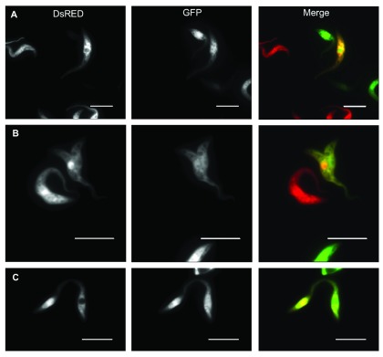
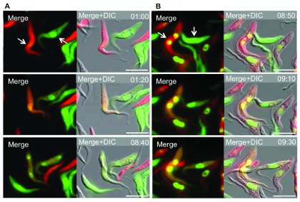
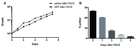
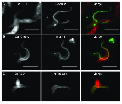
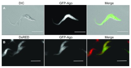
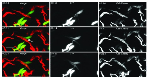
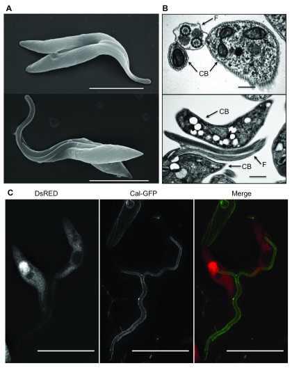
Diverse structures facilitate direct exchange of proteins between cells, including plasmadesmata in plants and tunnelling nanotubes in bacteria and higher eukaryotes. Here we describe a new mechanism of protein transfer, flagellar membrane fusion, in the unicellular parasite Trypanosoma brucei. When fluorescently tagged trypanosomes were co-cultured, a small proportion of double-positive cells were observed. The formation of double-positive cells was dependent on the presence of extracellular calcium and was enhanced by placing cells in medium supplemented with fresh bovine serum. Time-lapse microscopy revealed that double-positive cells arose by bidirectional protein exchange in the absence of nuclear transfer. Furthermore, super-resolution microscopy showed that this process occurred in ≤1 minute, the limit of temporal resolution in these experiments. Both cytoplasmic and membrane proteins could be transferred provided they gained access to the flagellum. Intriguingly, a component of the RNAi machinery (Argonaute) was able to move between cells, raising the possibility that small interfering RNAs are transported as cargo. Transmission electron microscopy showed that shared flagella contained two axonemes and two paraflagellar rods bounded by a single membrane. In some cases flagellar fusion was partial and interactions between cells were transient. In other cases fusion occurred along the entire length of the flagellum, was stable for several hours and might be irreversible. Fusion did not appear to be deleterious for cell function: paired cells were motile and could give rise to progeny while fused. The motile flagella of unicellular organisms are related to the sensory cilia of higher eukaryotes, raising the possibility that protein transfer between cells via cilia or flagella occurs more widely in nature.

Keywords: Trypanosoma; cell-cell communication; cilium; flagellum; lattice light sheet microscopy; membrane fusion; protein transfer; structured illumination microscopy.


Conflict of interest statement

Competing interests: The authors declare that they have no competing interests.

Figures

Figure 1.
Figure 1.. Fluorescence microscopy of co-cultured trypanosomes expressing DsRED or GFP.
A: Double-positive trypanosome after 24 hours co-culture (WT). B: Interacting double-positive trypanosome pair found after 24 hours co-culture (Δproc). C: Double-positive trypanosomes connected at their anterior ends (Δproc). The scale bar indicates 10μm. DsRed tends to accumulate in the nuclei of cells that synthesise it, probably because of its propensity to form tetramers.
Figure 2.
Figure 2.. Trypanosomes exchange proteins but not DNA in a contact-dependent manner.
Still images from time-lapse fluorescent microscopy of a mixed culture of wild type trypanosomes expressing Histone2B-GFP/cytosolic DsRED or Histone2B-GFP/cytosolic GFP. The time interval between images is indicated at the right corner of the image; the scale bar indicates 10μm; arrows indicate interacting cells. A: First image taken after one hour, the cells are clearly separate and only positive for one fluorescent protein in the cytoplasm. Second image 20 minutes later (1:20), cells are in contact and have become positive for DsRED and GFP. Third image, 7 hours and 20 minutes later (8:40), the cells have separated again. ( Video 1) B: First image taken after 8 hours 50 minutes, cells are only positive for one fluorescent protein in the cytoplasm. Second image, cells have become double-positive (09:10). Third image, 20 minutes later the cells have separated again (09:30). ( Video 2).
Figure 3.
Figure 3.. Double-positive cells lose one fluorescent protein over time.
A: Growth curve of Δproc yellow cells after FACS and Δproc GFP cells after FACS. B: Double-positive Δproc cells were enriched by FACS and the percentage of yellow cells was measured by flow cytometry at daily intervals.
Figure 4.
Figure 4.. Exchange of different fluorescently tagged proteins indicates involvement of the flagellum.
A: Interacting pair of trypanosomes expressing either DsRED (Δproc) or the GPI anchored surface protein EP-GFP (WT). B: Interacting pair of trypanosomes expressing the flagellar protein calflagin44-GFP (WT) or calflagin44-Cherry (WT). C: Interacting pair of trypanosomes expressing either cytoplasmic DsRED (Δproc) or the nucleoside transporter NT10-GFP (WT), which localises to the surface of the cell body, but not to the flagellum. The scale bar indicates 10μm.
Figure 5.
Figure 5.. GFP-tagged Ago1 partially localises to the flagellum and can be exchanged between cells.
A: Fluorescence microscopy of wild-type cells expressing GFP-Ago. B: Fluorescence microscopy of co-cultured wild-type cells expressing DsRED or GFP-Ago. Scale bars indicate 10μm.
Figure 6.
Figure 6.. Time-lapse microscopy shows a rapid exchange of cytoplasmic and flagellar proteins.
Still images from lattice light sheet fluorescence microscopy time-lapse video ( Video 3) of trypanosomes expressing either GFP (Δproc) or calflagin44-mCherry (WT). The time interval is indicated at the left upper corner of the image. The scale bar is 10μm. First image (00:10): cells are only positive for one fluorescent protein. Second image one minute later (00:11), calflagin44-mCherry is present on both cells, but GFP is detected only weakly in the second cell. Third image: two minutes later (00:13), GFP is equally distributed in both interacting cells.
Figure 7.
Figure 7.. High-resolution images of interacting trypanosomes.
A: Scanning electron microscopy of interacting trypanosomes (Δproc). The scale bar indicates 10μm. B: Transmission electron microscopy of fused flagella (Δproc). The scale bar indicates 0.5μm for the upper image and 1.5μm for the lower image; arrows indicate the cell-body (CB) and the flagellum (F). C: Structured illumination microscopy (SIM) of interacting trypanosomes with fused flagella. Trypanosomes were tagged with either DsRED (Δproc) or calflagin44-GFP (WT). The scale bar indicates 10μm.

Similar articles

Cited by

References

    1. Lucas WJ, Ham LK, Kim JY: Plasmodesmata - bridging the gap between neighboring plant cells. Trends Cell Biol. 2009;19(10):495–503. 10.1016/j.tcb.2009.07.003 - DOI - PubMed
    1. Goodenough DA, Paul DL: Gap junctions. Cold Spring Harb Perspect Biol. 2009;1(1):a002576. 10.1101/cshperspect.a002576 - DOI - PMC - PubMed
    1. Jedd G, Pieuchot L: Multiple modes for gatekeeping at fungal cell-to-cell channels. Mol Microbiol. 2012;86(6):1291–1294. 10.1111/mmi.12074 - DOI - PubMed
    1. Bloemendal S, Kück U: Cell-to-cell communication in plants, animals, and fungi: a comparative review. Naturwissenschaften. 2013;100(1):3–19. 10.1007/s00114-012-0988-z - DOI - PubMed
    1. Rustom A, Saffrich R, Markovic I, et al. : Nanotubular highways for intercellular organelle transport. Science. 2004;303(5660):1007–1010. 10.1126/science.1093133 - DOI - PubMed