. 2016 Jul;34(7):774-80.
doi: 10.1038/nbt.3563. Epub 2016 May 30.

A split horseradish peroxidase for the detection of intercellular protein-protein interactions and sensitive visualization of synapses

Affiliations

A split horseradish peroxidase for the detection of intercellular protein-protein interactions and sensitive visualization of synapses

Jeffrey D Martell et al. Nat Biotechnol. 2016 Jul.

Abstract

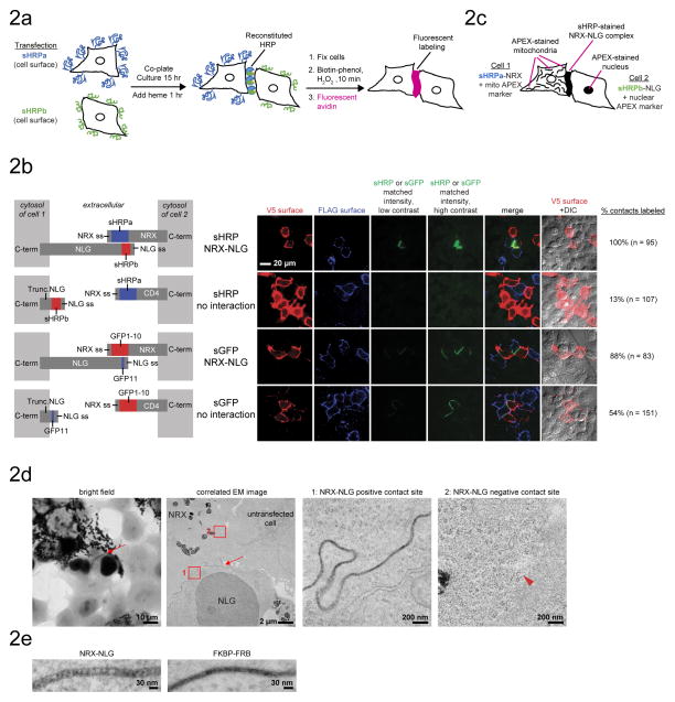
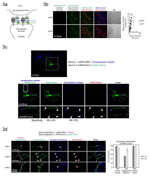
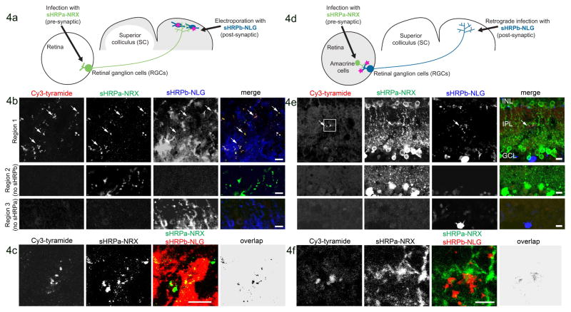
Intercellular protein-protein interactions (PPIs) enable communication between cells in diverse biological processes, including cell proliferation, immune responses, infection, and synaptic transmission, but they are challenging to visualize because existing techniques have insufficient sensitivity and/or specificity. Here we report a split horseradish peroxidase (sHRP) as a sensitive and specific tool for the detection of intercellular PPIs. The two sHRP fragments, engineered through screening of 17 cut sites in HRP followed by directed evolution, reconstitute into an active form when driven together by an intercellular PPI, producing bright fluorescence or contrast for electron microscopy. Fusing the sHRP fragments to the proteins neurexin (NRX) and neuroligin (NLG), which bind each other across the synaptic cleft, enabled sensitive visualization of synapses between specific sets of neurons, including two classes of synapses in the mouse visual system. sHRP should be widely applicable to studying mechanisms of communication between a variety of cell types.


Conflict of interest statement

COMPETING INTERESTS STATEMENT

Massachusetts Institute of Technology has filed a patent covering part of the information contained in this article.

Figures

Figure 1
Figure 1. Protein engineering of split HRP (sHRP)
A. Schematic overview of the split HRP reporter. Two inactive fragments of HRP reconstitute into an active complex capable of producing a variety of enzymatic reaction products, visible by multiple modalities. The nonfluorescent Amplex Red is converted to fluorescent resorufin. Biotin-phenol is converted to a reactive radical that becomes covalently attached to neighboring proteins. Tyramide signal amplification (TSA) substrates function analogously, except biotin is replaced with a fluorophore, such as Cy3. Diaminobenzidine (DAB) is converted to a colored and insoluble product that becomes electron-dense upon treatment with osmium tetroxide. B. Overview of sHRP protein engineering. Structure-guided cut-site screening was followed by two rounds of yeast display directed evolution. C. Overview of yeast display evolution of sHRPa (top) and sHRPb (bottom). D. Fluorescence activated cell sorting (FACS) of yeast displaying sHRP fragments at various stages of evolution. Aga1p and 2p are cell surface mating proteins. Re-amplified pools of yeast were labeled and analyzed under matched conditions (1 min biotin-phenol labeling). E. Crystal structure of HRP (PDB ID 1H5A). The sHRPa fragment is colored blue, and the sHRPb fragment is green. F. Amplex UltraRed live cell labeling followed by immunostaining of HEK293T cells expressing sHRP fragments in the ER lumen.
Figure 2
Figure 2. Intercellular reconstitution of sHRP for fluorescent and EM labeling
A. Scheme for intercellular sHRP reconstitution across HEK293T cells. B. Comparison of sHRP and split GFP (sGFP),,. Split HRP produces brighter fluorescence than split GFP, and its activity is dependent on a protein-protein interaction. The two pools of cells were visualized by surface immunostaining of N-terminal V5 and FLAG epitope tags. V5 and FLAG intensities are normalized between the two sHRP experiments and between the two sGFP experiments, but not across all 4 conditions. Surface expression levels of sHRP constructs and sGFP constructs were similar. Construct designs are shown at left. “ss” is signal sequence. C. Scheme for EM staining of HEK293T intercellular contacts by sHRP. APEX co-transfection markers provide contrast for both light microscopy and EM. D. EM imaging of the intercellular NRX-NLG protein-protein interaction using sHRP (as in C). Arrow points to contact site stained by sHRP. Arrowhead points to a contact site not stained by sHRP. The third and fourth images are zooms of boxed regions in second image. E. High magnification EM of sHRP staining generated by different protein-protein interactions.
Figure 3
Figure 3. Synapse detection in cultured neurons using sHRP
A. Scheme for reconstitution of sHRP by the trans-synaptic NRX-NLG interaction in the neuronal synaptic cleft. sHRP constructs are expressed under the synapsin promoter to minimize overexpression artifacts. B. Comparison of sHRP and split GFP,, for synapse detection. Constructs with synapsin promoter were introduced into separate neuron populations using a two-step transfection procedure. Confocal images are shown at left, and quantitation of maximum signal/noise as a function of contact site area is shown at right. Plots present >70 contact sites across >5 fields of view for each condition. The sHRP-NRX neurons and the sGFP-NLG neurons were marked by a Tomato co-transfection marker, and the sHRP-NLG neurons were marked by a Venus co-transfection marker. sGFP-NRX neurons were detected by anti-V5 staining (AlexaFluor 647 readout). Intensity scales are not normalized for the transfection markers. C. sHRP labeling is localized to synapses. Confocal fluorescence imaging of sHRP (synapsin promoter) with respect to pre- and post-synaptic markers, synaptophysin-mApple and Homer-Venus, respectively. Arrowheads point to sHRP labeling sites. Specificity is calculated as the fraction of sHRP puncta that overlap with each marker. Values are the mean +/− std dev of 3 independent experiments. D. Confocal fluorescence imaging of sHRP (synapsin promoter) with respect to endogenous Bassoon, a pre-synaptic marker. The graph presents the % overlap of sHRP puncta. Data in bar graph represent the mean +/− std dev of 3 independent experiments. Experiments were performed at either 16 days in vitro (DIV) or 20 days in vitro.
Figure 4
Figure 4. Detection of reconstituted sHRP in vivo
A. Scheme for detection of anterograde contacts between RGCs and neurons of the SC. B. Four weeks after mouse transduction as in (A), the SC was removed by dissection, incubated with heme, fixed, sectioned, and labeled with Cy3-tyramide, a fluorescent sHRP substrate. sHRPa-NRX and sHRPb-NLG expression were detected by staining with anti-HRP and anti-NLG antibodies, respectively (anti-HRP does not recognize sHRPb). The second and third rows show tissue regions lacking sHRPb or sHRPa, respectively. Arrows point to sHRP-stained terminals. C. Labeled SC from a different animal. “Overlap” is the calculated overlap between sHRPa and sHRPb channels. This experiment was repeated 8 times with similar results. D. Scheme for detection of retrograde contacts between RGCs and amacrine cells in the mouse retina. sHRPa-NRX expression was restricted to amacrine cells via use of a Cre line (see Methods). E. One week after transduction as in (D), the retina was dissected and prepared as in (B). Second and third rows show tissue regions lacking sHRPb or sHRPa expression, respectively. Arrow points to sHRP staining. Box indicates the region shown in (F). F. Zoom region indicated by the box in (E). All scale bars, 10 μm.

References

    1. Kim SA, Tai CY, Mok LP, Mosser EA, Schuman EM. Calcium-dependent dynamics of cadherin interactions at cell–cell junctions. Proceedings of the National Academy of Sciences. 2011;108:9857–9862. - PMC - PubMed
    1. Feinberg EH, et al. GFP Reconstitution Across Synaptic Partners (GRASP) defines cell contacts and synapses in living nervous systems. Neuron. 2008;57:353–363. - PubMed
    1. Liu DS, Loh KH, Lam SS, White KA, Ting AY. Imaging trans-cellular neurexin-neuroligin interactions by enzymatic probe ligation. PloS one. 2013;8:e52823. - PMC - PubMed
    1. Craig AM, Kang Y. Neurexin–neuroligin signaling in synapse development. Current opinion in neurobiology. 2007;17:43–52. - PMC - PubMed
    1. Michnick SW, Ear PH, Manderson EN, Remy I, Stefan E. Universal strategies in research and drug discovery based on protein-fragment complementation assays. Nature reviews Drug discovery. 2007;6:569–582. - PubMed

Publication types

MeSH terms

Substances