

Heterogeneous Pathology of Melasma and Its Clinical Implications
- PMID: 27240341
- PMCID: PMC4926358
- DOI: 10.3390/ijms17060824
Heterogeneous Pathology of Melasma and Its Clinical Implications
Abstract
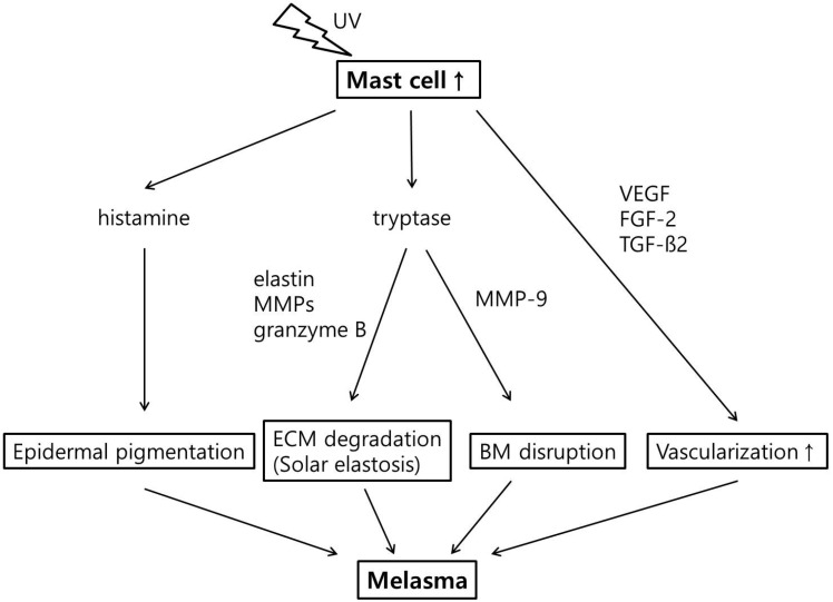
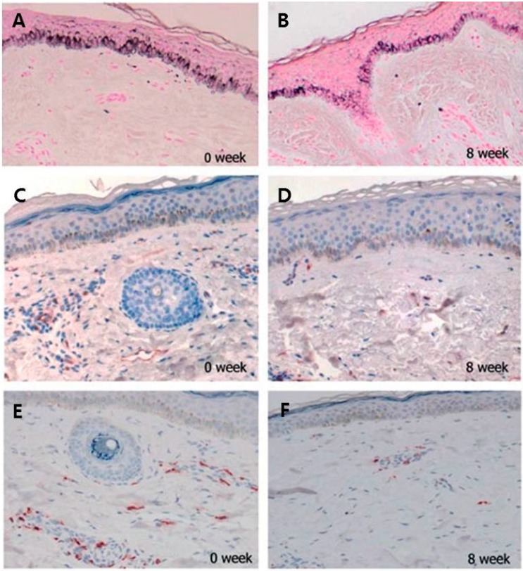
Melasma is a commonly acquired hypermelanosis that affects sun-exposed areas of the skin, with frequent facial involvement. Its histologic manifestations are evident in the epidermis, extracellular matrix, and dermis. In addition to epidermal pigmentation, pathologic findings of melasma include extracellular matrix abnormality, especially solar elastosis. The disrupted basement membrane has been described in melasma with variable incidences. In the dermis, an increase in vascularity and an increase in the number of mast cells were observed, indicating that dermal factors have critical roles in the pathogenesis of melasma, despite the fact that melasma is characterized by epidermal hyperpigmentation. This review discusses such histologic characteristics of melasma, with consideration to their implications for melasma treatment.
Keywords: basement membrane; histopathology; mast cells; melasma; photoaging; vascularization.
Figures
References
-
- Kwon S.H., Park K.C. Melasma and common pigmentary dermatoses in Asian individuals and an overview of their treatment. J. Clin. Investig. Dermatol. 2014;2:e8.
-
- Ortonne J.P., Arellano I., Berneburg M., Cestari T., Chan H., Grimes P., Hexsel D., Im S., Lim J., Lui H., et al. A global survey of the role of ultraviolet radiation and hormonal influences in the development of melasma. J. Eur. Acad. Dermatol. Venereol. 2009;23:1254–1262. doi: 10.1111/j.1468-3083.2009.03295.x. - DOI - PubMed
Publication types
MeSH terms
Substances
LinkOut - more resources
Full Text Sources
Other Literature Sources
