Review
. 2016 May 28;17(6):842.
doi: 10.3390/ijms17060842.

MicroRNAs: Key Regulators in the Central Nervous System and Their Implication in Neurological Diseases

Affiliations
Review

MicroRNAs: Key Regulators in the Central Nervous System and Their Implication in Neurological Diseases

Dan-Dan Cao et al. Int J Mol Sci. .

Abstract

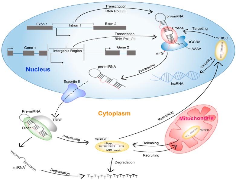
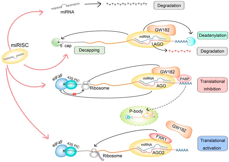
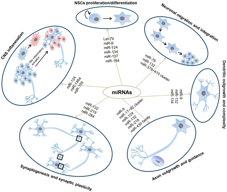
MicroRNAs (miRNAs) are a class of small, well-conserved noncoding RNAs that regulate gene expression post-transcriptionally. They have been demonstrated to regulate a lot of biological pathways and cellular functions. Many miRNAs are dynamically regulated during central nervous system (CNS) development and are spatially expressed in adult brain indicating their essential roles in neural development and function. In addition, accumulating evidence strongly suggests that dysfunction of miRNAs contributes to neurological diseases. These observations, together with their gene regulation property, implicated miRNAs to be the key regulators in the complex genetic network of the CNS. In this review, we first focus on the ways through which miRNAs exert the regulatory function and how miRNAs are regulated in the CNS. We then summarize recent findings that highlight the versatile roles of miRNAs in normal CNS physiology and their association with several types of neurological diseases. Subsequently we discuss the limitations of miRNAs research based on current studies as well as the potential therapeutic applications and challenges of miRNAs in neurological disorders. We endeavor to provide an updated description of the regulatory roles of miRNAs in normal CNS functions and pathogenesis of neurological diseases.

Keywords: CNS development; microRNA; neurogenesis; neurological diseases; regulation.


Figures

Figure 1
Figure 1
miRNA biogenesis and action. Pri-miRNA is typically transcribed from intron or intergenic region by polymerase II or polymerase III (Pol II or Pol III). In the nucleus the pri-miRNA is recognized and cleaved by Drosha and its partner DGCR8 to generate the ~70 nucleotides of pre-miRNA. The nuclear export of pre-miRNA molecules into the cytoplasm is mediated by Exportin 5 (XPO5) where they are further processed by Dicer with the aid of TAR RNA binding protein (TRBP) to generate the duplex of miRNA:miRNA*. Generally, the miRNA* is released to be degraded, while the miRNA is loaded into Agronaute (AGO) protein to form the miRNA-induced silencing complexes (miRISC) which could regulate the gene expression post-transcriptionally. When the target mRNAs are unavailable, the miRNA would also decay after being released from the miRISC. In some cases, the miRISC is recruited to mitochondria or relocated to nucleus, at which it can target diverse targets including pri-miRNAs and long non-coding RNAs (lncRNAs).
Figure 2
Figure 2
miRNA regulatory mechanisms. When the target mRNAs are not available, or not highly complementary to miRNAs, although rare in mammals, the miRNAs would be degraded. In most of cases, the miRISC, which generally constitute of miRNA, AGO protein and miRNA-induced silencing complexes (GW182), would target the 3′-UTR of mRNA. The imperfect complementary bet-ween miRNA and the 3′-UTR of mRNA would destabilize the 3′ poly(A) tail and 5′ cap of the mRNA and lead to degradation. Besides, the binding of miRISC with auxiliary protein poly(A)-binding protein (PABP) to mRNA could cause the translational inhibition via repressing activity of the eIF4F complex and the 43S pre-initiation complex (PIC). The dynamics structure P-body (processing body) could also sequester the target mRNA from the translation process. In a few cases, the translation of the mRNA in ribosome is promoted instead of inhibited by the miRISC. The red line represents major pathways of miRISC, the dark line represents interaction between different components, and the dotted line represents alternative way of translation inhibition processed by P-body.
Figure 3
Figure 3
Roles of miRNAs during neuronal development. Listed are miRNAs covered in this review that are functional in each stage of neuronal development. Red indicates activated microglia. Black square indicates synapses.

References

    1. Ramalingam P., Palanichamy J.K., Singh A., Das P., Bhagat M., Kassab M.A., Sinha S., Chattopadhyay P. Biogenesis of intronic miRNAs located in clusters by independent transcription and alternative splicing. RNA. 2014;20:76–87. doi: 10.1261/rna.041814.113. - DOI - PMC - PubMed
    1. Lee Y., Kim M., Han J., Yeom K.H., Lee S., Baek S.H., Kim V.N. MicroRNA genes are transcribed by RNA polymerase II. EMBO J. 2004;23:4051–4060. doi: 10.1038/sj.emboj.7600385. - DOI - PMC - PubMed
    1. Borchert G.M., Lanier W., Davidson B.L. RNA polymerase III transcribes human microRNAs. Nat. Struct. Mol. Biol. 2006;13:1097–1101. doi: 10.1038/nsmb1167. - DOI - PubMed
    1. Han J., Lee Y., Yeom K.H., Kim Y.K., Jin H., Kim V.N. The drosha-DGCR8 complex in primary microRNA processing. Genes Dev. 2004;18:3016–3027. doi: 10.1101/gad.1262504. - DOI - PMC - PubMed
    1. Yi R., Qin Y., Macara I.G., Cullen B.R. Exportin-5 mediates the nuclear export of pre-microRNAs and short hairpin rnas. Genes Dev. 2003;17:3011–3016. doi: 10.1101/gad.1158803. - DOI - PMC - PubMed

MeSH terms

LinkOut - more resources