

The measurement of the normal thorax using the Haller index methodology at multiple vertebral levels
- PMID: 27240848
- PMCID: PMC5013057
- DOI: 10.1111/joa.12499
The measurement of the normal thorax using the Haller index methodology at multiple vertebral levels
Abstract
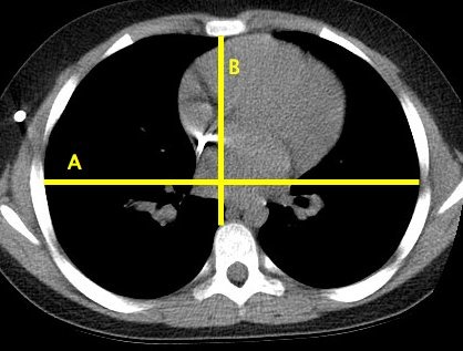
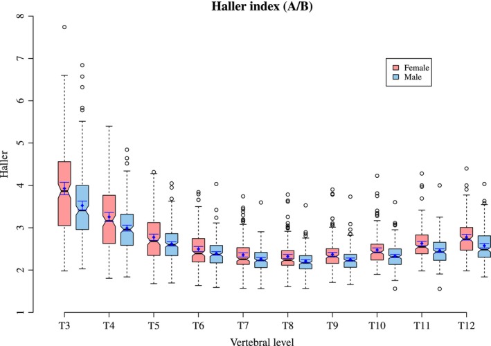
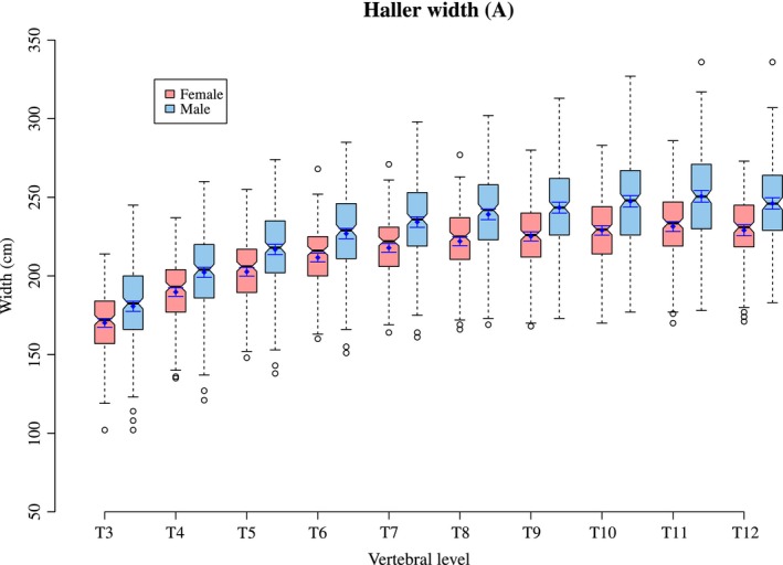
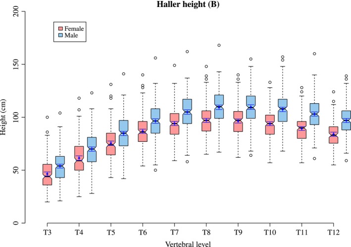
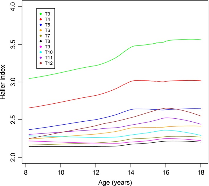
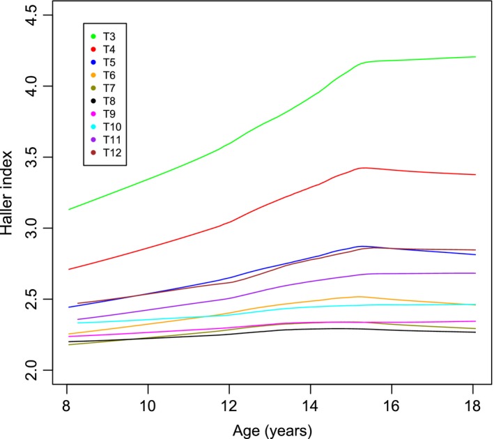
The Haller index is a ratio of thoracic width and height, measured from an axial CT image and used to describe the internal dimensions of the thoracic cage. Although the Haller index for a normal thorax has been established (Haller et al. 1987; Daunt et al. 2004), this is only at one undefined vertebral level in the thorax. What is not clear is how the Haller index describes the thorax at every vertebral level in the absence of sternal deformity, or how this is affected by age. This paper documents the shape of the thorax using the Haller index calculated from the thoracic width and height at all vertebral levels of the thorax between 8 and 18 years of age. The Haller Index changes with vertebral level, with the largest ratio seen in the most cranial levels of the thorax. Increasing age alters the shape of the thorax, with the most cranial vertebral levels having a greater Haller index over the mid thorax, which does not change. A slight increase is seen in the more caudal vertebral levels. These data highlight that a 'one size fits all' rule for chest width and depth ratio at all ages and all thoracic levels is not appropriate. The normal range for width to height ratio should be based on a patient's age and vertebral level.
Keywords: CT; Haller index; chest wall; paediatric; pectus excavatum; sternum; thoracic cage.
© 2016 Anatomical Society.
Figures
References
-
- Albertal M, Vallejos J, Bellia G, et al. (2013) Changes in chest compression indexes with breathing underestimate surgical candidacy in patients with pectus excavatum: a computed tomography pilot study. J Pediatr Surg 48, 2011–2016. - PubMed
-
- Birkemeier K, Podberesky D, Salisbury S, et al. (2011) Breathe in… breathe out… stop breathing: does phase of respiration affect the Haller index in patients with pectus excavatum? AJR Am J Roentgenol 197, W934–W939. - PubMed
-
- Daunt S, Cohen J, Miller S (2004) Age‐related normal ranges for the Haller index in children. Pediatr Radiol 34, 326–330. - PubMed
-
- Haller J, Kramer S, Leitman S (1987) Use of CT scans in selection of patients for pectus excavatum surgery: a preliminary report. J Pediatr Surg 22, 904–906. - PubMed
-
- Hong J‐Y, Suh S‐W, Easwar T, et al. (2011) Evaluation of the three‐dimensional deformities in scoliosis surgery with computed tomography: efficacy and relationship with clinical outcomes. Spine 36, E1259–E1265. - PubMed
MeSH terms
LinkOut - more resources
Full Text Sources
Other Literature Sources
