. 2016 May 31:6:26822.
doi: 10.1038/srep26822.

Analysis of the dynamic co-expression network of heart regeneration in the zebrafish

Affiliations

Analysis of the dynamic co-expression network of heart regeneration in the zebrafish

Sophie Rodius et al. Sci Rep. .

Abstract

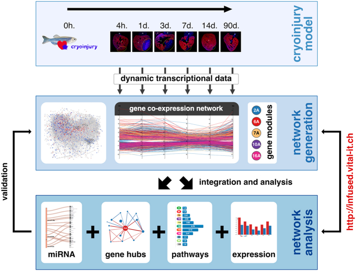
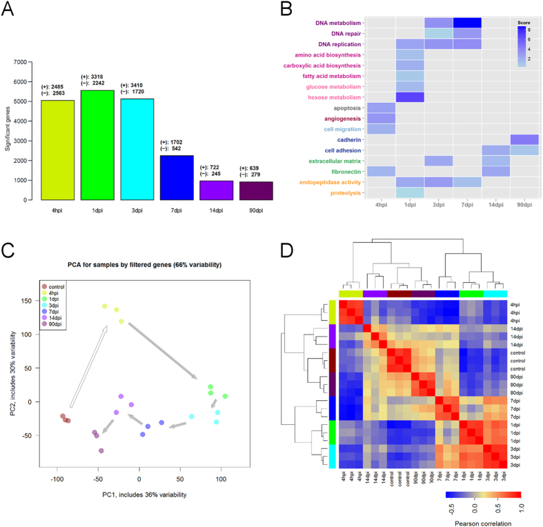
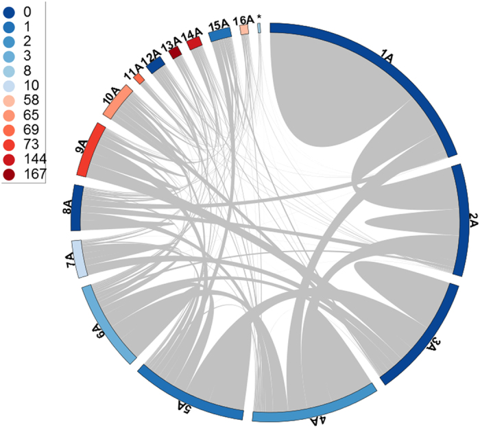
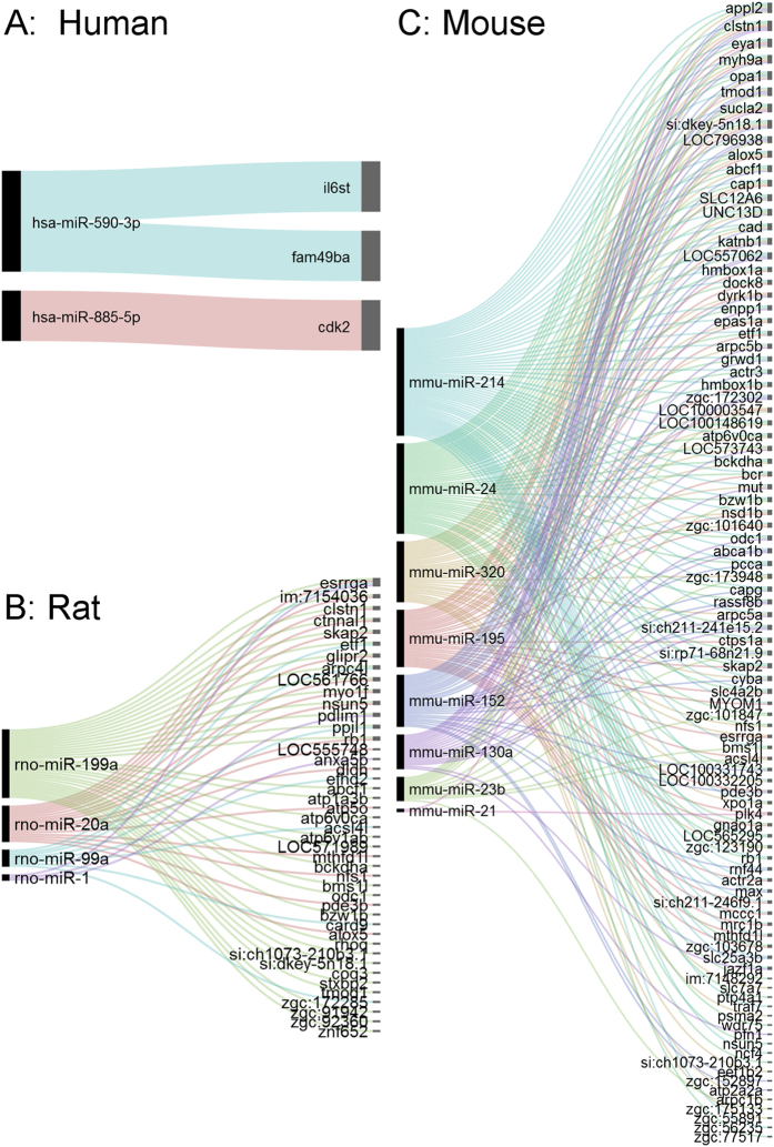
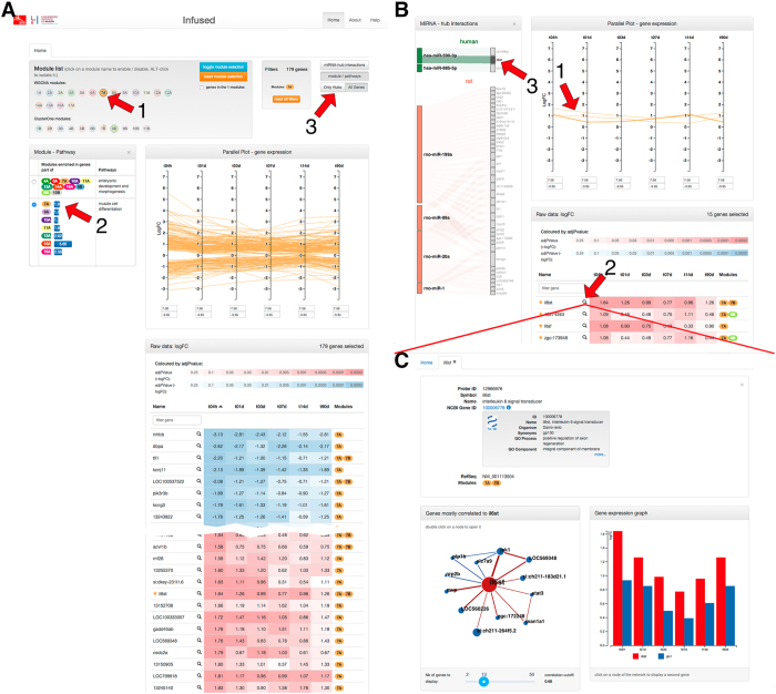
The zebrafish has the capacity to regenerate its heart after severe injury. While the function of a few genes during this process has been studied, we are far from fully understanding how genes interact to coordinate heart regeneration. To enable systematic insights into this phenomenon, we generated and integrated a dynamic co-expression network of heart regeneration in the zebrafish and linked systems-level properties to the underlying molecular events. Across multiple post-injury time points, the network displays topological attributes of biological relevance. We show that regeneration steps are mediated by modules of transcriptionally coordinated genes, and by genes acting as network hubs. We also established direct associations between hubs and validated drivers of heart regeneration with murine and human orthologs. The resulting models and interactive analysis tools are available at http://infused.vital-it.ch. Using a worked example, we demonstrate the usefulness of this unique open resource for hypothesis generation and in silico screening for genes involved in heart regeneration.


Conflict of interest statement

The authors declare no competing financial interests.

Figures

Figure 1
Figure 1. Discovery and resource development framework.
Gene expression was measured and co-expression networks generated from control and injured zebrafish hearts at different times. Network structure analyses were implemented and their relations to biological function were determined, and top predictions were further investigated. This includes an in silico validation phase, which involved the establishment of association between the detected hubs (and modules) and independent external biological information from zebrafish and mammals (sources are indicated in box “network analysis”). A Web-based interactive resource is provided to enable the analysis, integration and visualization of these datasets. The zebrafish drawing was adapted from (http://bit.ly/1PL92vj) and is licensed under the Attribution-Share-Alike 3.0 Unported license (terms can be found at http://bit.ly/1pawxfE).
Figure 2
Figure 2. The different stages of heart regeneration in the zebrafish.
Sagittal sections of adult zebrafish heart: anterior is towards the top and ventral towards the right. Hearts were cryoinjured and recovered at the indicated time points post-injury, and compared to healthy hearts from control fish. Sections were immunostained for tropomyosin in red, or TUNEL in green (nuclei are stained with DAPI in blue). Fibrosis was monitored by Masson-Goldner trichrome staining: healthy myocardial tissue (in red) and fibrotic areas (in blue). A: atrium; B: bulbus arteriosus; V: ventricle; hpi: hours post-injury; dpi: days post-injury. The injured area is indicated by a dotted circle. Scale bar is 100 μm.
Figure 3
Figure 3. Significant changes in gene expression during heart regeneration.
(A) Numbers of DEGs at each time point. (B) Summary of statistically enriched functional terms of DEGs at each time point. (C) Visualization of control and post-injury samples based on PCA (genes with FDR < 0.001). (D) Hierarchical clustering of samples based on gene expression data (genes with FDR < 0.001).
Figure 4
Figure 4. Modular architecture of the gene co-expression network in the zebrafish heart regeneration.
Circular plots of modules detected with WGCNA: Internal links (grey) represent the intra- and inter-module connectivity, whereas the color of the outer bar represents the number of functional terms significantly enriched in a given module (Fig. 5). Numbers shown next to the colored circles (left panel) indicate the number of functional terms associated with each color in the plot.
Figure 5
Figure 5. Summary of functional enrichments of modules.
Dotted line represents threshold of statistical significance at FDR = 0.05 (Supplementary Methods).
Figure 6
Figure 6. Network hubs are functionally important for heart regeneration in mammals.
It offers an overall view of biologically important associations between hubs and miRs known to be drivers of regeneration in mammals. Higher resolution views, including their integration with other biological information, are provided on the website. (A) miR-hub interactions in humans. (B) miR-hub interactions in the rat. (C) miR-hub interactions in the mouse. Lines are used to indicate miR-target interactions, and are colored to group miR-specific interactions.
Figure 7
Figure 7. The web platform enables to explore the resource content; example of il6st.
Select a module to explore (A-1, module 7A). Parallel coordinate plots as well as colored table of gene expression values (log2 fold change) present the profile and significance of gene expression changes in response to cryoinjury. Hub genes are marked with a star in the table. The functional categories enriched in this module are listed in (A-2). Panel (B) presents a restricted view on hub genes (available by clicking on A-3). (B-3) reveals a putative link between il6st and miR-885-5p in human. A detailed view on il6st gene (C) is displayed after clicking on (B-2). A summary of the NCBI gene record of that gene is available. (C-1) displays the network of genes most correlated to il6st. By clicking on any of the nodes (Jak1 in this example), the expression fold change of the gene is displayed side-by-side against il6st (C-2).

References

    1. Kikuchi K, Poss KD. Cardiac Regenerative Capacity and Mechanisms. Annu. Rev. Cell Dev. Biol. 2012;28:719–741. doi: 10.1146/annurev-cellbio-101011-155739. - DOI - PMC - PubMed
    1. Gemberling M, Bailey TJ, Hyde DR, Poss KD. The zebrafish as a model for complex tissue regeneration. Trends Genet. 2013;29:611–620. doi: 10.1016/j.tig.2013.07.003. - DOI - PMC - PubMed
    1. Poss KD, Wilson LG, Keating MT. Heart regeneration in zebrafish. Science. 2002;298:2188–90. doi: 10.1126/science.1077857. - DOI - PubMed
    1. Schnabel K, Wu C-C, Kurth T, Weidinger G. Regeneration of cryoinjury induced necrotic heart lesions in zebrafish is associated with epicardial activation and cardiomyocyte proliferation. Plos One. 2011;6:e18503. doi: 10.1371/journal.pone.0018503. - DOI - PMC - PubMed
    1. Gonzalez-Rosa JM, Martin V, Peralta M, Torres M, Mercader N. Extensive scar formation and regression during heart regeneration after cryoinjury in zebrafish. Development. 2011;138:1663–1674. doi: 10.1242/dev.060897. - DOI - PubMed

Publication types

Substances