Review
. 2016 Sep;13(9):1868-77.
doi: 10.1016/j.hrthm.2016.05.026. Epub 2016 May 27.

Ablating atrial fibrillation: A translational science perspective for clinicians

Affiliations
Review

Ablating atrial fibrillation: A translational science perspective for clinicians

James N Weiss et al. Heart Rhythm. 2016 Sep.

Abstract

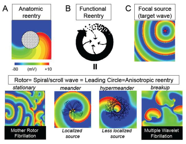
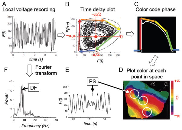
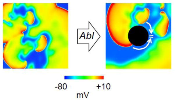
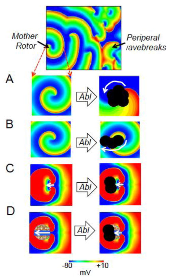
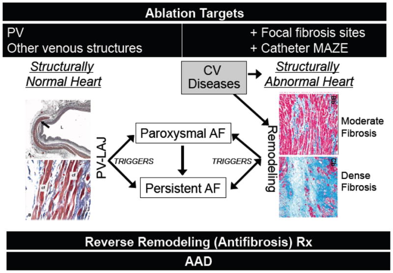
Although considerable progress has been made in developing ablation approaches to cure atrial fibrillation (AF), outcomes are still suboptimal, especially for persistent and long-lasting persistent AF. In this topical review, we review the arrhythmia mechanisms, both reentrant and nonreentrant, that are potentially relevant to human AF at various stages/settings. We describe arrhythmia mapping techniques used to distinguish between the different mechanisms, with a particular focus on the detection of rotors. We discuss which arrhythmia mechanisms are likely to respond to ablation, and the challenges and prospects for improving upon current ablation strategies to achieve better outcomes.

Keywords: Ablation; Arrhythmia; Atrial fibrillation; Automaticity; Fibrosis; Reentry; Rotor; Triggered activity.


Conflict of interest statement

None

Figures

Figure 1
Figure 1. Basic arrhythmia mechanisms relevant to fibrillation
A. Anatomic reentry (A), in which the wavefront rotates around an inexcitable anatomic obstacle. B. Functional reentry (leading circle=anisotropic=spiral/scroll wave), in which a rotor rotates around a core of excitable, but unexcited, tissue. Depending on electrophysiological characteristics of the tissue, the rotor can be stable (lower left panel) with peripheral wavebreaks (fibrillatory conduction block) if the surrounding tissue has a longer refractory period, meandering (lower left middle panel), hypermeandering (lower middle right panel), or in an unstable breakup regime (lower right panel). A stable or meandering rotor with peripheral wavebreak is equivalent to Mother Rotor Fibrillation, whereas spiral wave break-up is equivalent to Multiple Wavelet Fibrillation. C. Focal sources due to automaticity or EAD- or DAD-mediated triggered activity produce a target wave pattern of concentric wavefronts. Except for the middle upper panel, all other panels show color-coded voltage (blue repolarized, red-green depolarized) voltage snapshots. The temporal trajectories of the rotor cores are shown in black lines for the meandering and hypermeandering rotors. Panel B was adapted from Allessie et al with permission.
Fig. 2
Fig. 2. Principles of phase-mapping and dominant frequency (DF) determination
A. Optically-recorded trace of voltage fluorescence (F(t)) from a point on the surface of the heart during a tachyarrhythmia. B. Transformation of the voltage trace in A to a time delay plot, in which F(t) at time t is plotted against F(t+τ), i.e. the voltage fluorescence at a later time t+τ. When τ is chosen properly, this produces a circular pattern. The phase at any given time is then defined as the angle (from -π to π) of a line drawn from the center of the circle (red dot) to the position on the circular pattern at that point in time (analogous to time on a clockface). C. The angles are color-coded corresponding to the different phases of action potential recorded by the voltage fluorescence at that location. D. A snapshot of the color (phase) at each location over the surface of the heart at a given time point generates a phase-map. E. The rotation centers (cores) of rotors have small voltage oscillations (corresponding to low amplitude double potentials on extracellular electrograms). Since their phase is indeterminate, they are called phase singularities (PS). They appear as the rotation centers of color wheels on the phase map (white circles in D). In contrast, a focal source emanating from automaticity or triggered activity appears as a target wave of colors (phases), analogous to Fig. 1C. F. Alternatively, the fluorescence trace in A can be converted from the time domain to the frequency domain using a Fourier transform. The largest peak is called the dominant frequency (DF). Panels A, B, D–F were adapted from Gray et al , with permission.
Fig. 3
Fig. 3. Focal ablation lesions accelerate termination of MW Fibrillation
Simulation in 2D tissue (10×10 cm) illustrating MW Fibrillation before (left) and after (right) an obstacle (ablation lesion, black circle) is created. The obstacle anchors two of the rotor tips causing them to collide and annihilate (arrows), while the remaining rotor tips self-extinguish at the tissue borders, terminating MW Fibrillation. Adapted from Qu et al. , with permission.
Fig. 4
Fig. 4. Ablatability of MR Fibrillation (upper panel)
A. Ablation (Abl) converts the Mother Rotor from functional reentry (left panel) to slower anatomic reentry around the ablation lesion (right panel, red arrow). B. Ablation creates a lesion extending from the Mother Rotor core to a border, interrupting the reentrant circuit (left panel). C. Ablation creates a lesion in the central common pathway of a figure of eight Mother Rotor. D. The Mother Rotor is not true functional reentry, but anatomic micro-reentry dependent on slow conduction through a channel (right panel), which is interrupted by the ablation lesion (left panel).
Fig. 5
Fig. 5. Conventional ablation targets in structurally normal and abnormal hearts
Left panels show histology of PV sleeves near the PV-LA junction (PV-LAJ), with cardiac myocytes strands (red) separated by collagen bundles (blue) and vascular tissue, from which triggers emerge to initiate AF. Right panels show histology of remodeled atrial tissue, with myocyte strands (red) interdigitated with collagen bundles (upper) and dense fibrosis (lower), promoting both triggers and slow conduction. Both CV diseases and persistent AF promote remodeling. Histology panels reproduced with permission from and . AAD, antiarrhythmic drugs

References

    1. Haissaguerre M, Jais P, Shah DC, Takahashi A, Hocini M, Quiniou G, Garrigue S, Le Mouroux A, Le Metayer P, Clementy J. Spontaneous initiation of atrial fibrillation by ectopic beats originating in the pulmonary veins. N Engl J Med. 1998;339:659–666. - PubMed
    1. Verma A, Jiang CY, Betts TR, et al. Approaches to catheter ablation for persistent atrial fibrillation. N Engl J Med. 2015;372:1812–1822. - PubMed
    1. Narayan SM, Krummen DE, Shivkumar K, Clopton P, Rappel WJ, Miller JM. Treatment of atrial fibrillation by the ablation of localized sources: CONFIRM (Conventional Ablation for Atrial Fibrillation With or Without Focal Impulse and Rotor Modulation) trial. J Am Coll Cardiol. 2012;60:628–636. - PMC - PubMed
    1. Narayan SM, Baykaner T, Clopton P, Schricker A, Lalani GG, Krummen DE, Shivkumar K, Miller JM. Ablation of rotor and focal sources reduces late recurrence of atrial fibrillation compared with trigger ablation alone: extended follow-up of the CONFIRM trial (Conventional Ablation for Atrial Fibrillation With or Without Focal Impulse and Rotor Modulation) Journal of the American College of Cardiology. 2014;63:1761–1768. - PMC - PubMed
    1. Miller JM, Kowal RC, Swarup V, et al. Initial independent outcomes from focal impulse and rotor modulation ablation for atrial fibrillation: multicenter FIRM registry. J Cardiovasc Electrophysiol. 2014;25:921–929. - PMC - PubMed

MeSH terms