

A highly soluble, non-phototoxic, non-fluorescent blebbistatin derivative
- PMID: 27241904
- PMCID: PMC4886532
- DOI: 10.1038/srep26141
A highly soluble, non-phototoxic, non-fluorescent blebbistatin derivative
Abstract
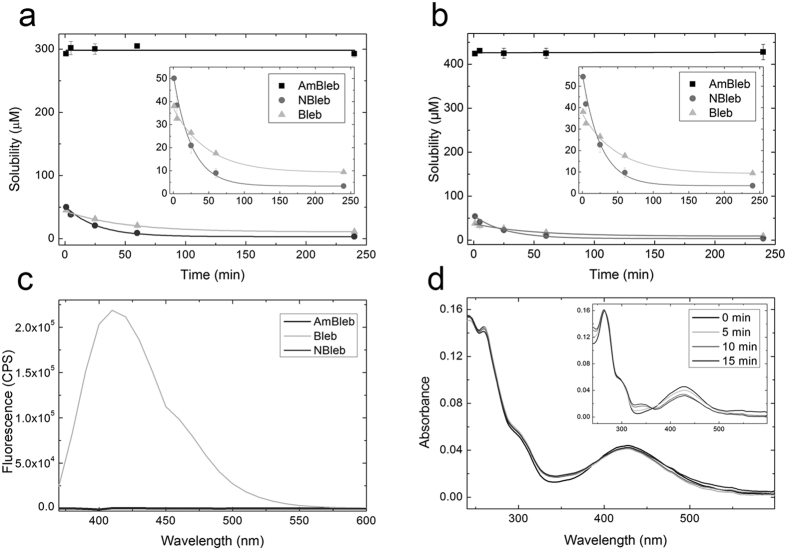
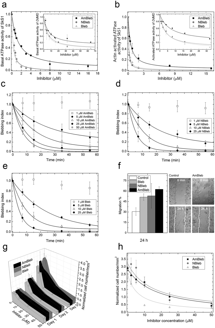
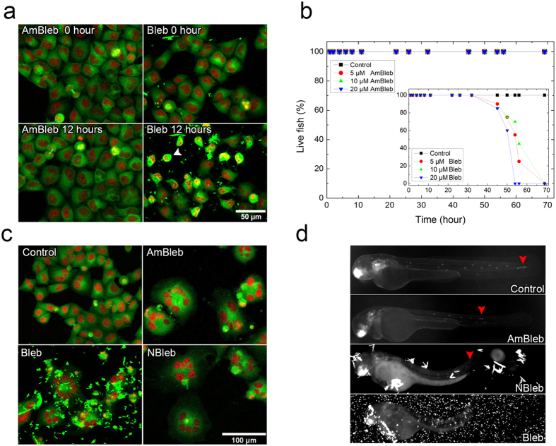
Blebbistatin is a commonly used molecular tool for the specific inhibition of various myosin II isoforms both in vitro and in vivo. Despite its popularity, the use of blebbistatin is hindered by its poor water-solubility (below 10 micromolar in aqueous buffer) and blue-light sensitivity, resulting in the photoconversion of the molecule, causing severe cellular phototoxicity in addition to its cytotoxicity. Furthermore, blebbistatin forms insoluble aggregates in water-based media above 10 micromolar with extremely high fluorescence and also high adherence to different types of surfaces, which biases its experimental usage. Here, we report a highly soluble (440 micromolar in aqueous buffer), non-fluorescent and photostable C15 amino-substituted derivative of blebbistatin, called para-aminoblebbistatin. Importantly, it is neither photo- nor cytotoxic, as demonstrated on HeLa cells and zebrafish embryos. Additionally, para-aminoblebbistatin bears similar myosin II inhibitory properties to blebbistatin or para-nitroblebbistatin (not to be confused with the C7 substituted nitroblebbistatin), tested on rabbit skeletal muscle myosin S1 and on M2 and HeLa cells. Due to its drastically improved solubility and photochemical feature, as well as lack of photo- or cytotoxicity, para-aminoblebbistatin may become a feasible replacement for blebbistatin, especially at applications when high concentrations of the inhibitor or blue light irradiation is required.
Conflict of interest statement
Patent application number: EP16153446, A.M.C. (corresponding author) is an owner in Optopharma Ltd.
Figures
Similar articles
-
para-Nitroblebbistatin, the non-cytotoxic and photostable myosin II inhibitor.Angew Chem Int Ed Engl. 2014 Jul 28;53(31):8211-5. doi: 10.1002/anie.201403540. Epub 2014 Jun 20. Angew Chem Int Ed Engl. 2014. PMID: 24954740
-
Discovery of (S)-3'-hydroxyblebbistatin and (S)-3'-aminoblebbistatin: polar myosin II inhibitors with superior research tool properties.Org Biomol Chem. 2017 Mar 1;15(9):2104-2118. doi: 10.1039/c7ob00006e. Org Biomol Chem. 2017. PMID: 28220174
-
Improved Inhibitory and Absorption, Distribution, Metabolism, Excretion, and Toxicology (ADMET) Properties of Blebbistatin Derivatives Indicate That Blebbistatin Scaffold Is Ideal for drug Development Targeting Myosin-2.J Pharmacol Exp Ther. 2021 Mar;376(3):358-373. doi: 10.1124/jpet.120.000167. Epub 2021 Jan 19. J Pharmacol Exp Ther. 2021. PMID: 33468641
-
Medicinal Chemistry and Use of Myosin II Inhibitor ( S)-Blebbistatin and Its Derivatives.J Med Chem. 2018 Nov 8;61(21):9410-9428. doi: 10.1021/acs.jmedchem.8b00503. Epub 2018 Jun 21. J Med Chem. 2018. PMID: 29878759 Review.
-
Targeting Myosin by Blebbistatin Derivatives: Optimization and Pharmacological Potential.Trends Biochem Sci. 2018 Sep;43(9):700-713. doi: 10.1016/j.tibs.2018.06.006. Epub 2018 Jul 26. Trends Biochem Sci. 2018. PMID: 30057142 Review.
Cited by
-
The role of actin and myosin in antigen extraction by B lymphocytes.Semin Cell Dev Biol. 2020 Jun;102:90-104. doi: 10.1016/j.semcdb.2019.10.017. Epub 2019 Dec 18. Semin Cell Dev Biol. 2020. PMID: 31862219 Free PMC article. Review.
-
Cytokinetic contractile ring structural progression in an early embryo: positioning of scaffolding proteins, recruitment of α-actinin, and effects of myosin II inhibition.Front Cell Dev Biol. 2024 Sep 27;12:1483345. doi: 10.3389/fcell.2024.1483345. eCollection 2024. Front Cell Dev Biol. 2024. PMID: 39398481 Free PMC article.
-
Knocking Out Non-muscle Myosin II in Retinal Ganglion Cells Promotes Long-Distance Optic Nerve Regeneration.Cell Rep. 2020 Apr 21;31(3):107537. doi: 10.1016/j.celrep.2020.107537. Cell Rep. 2020. PMID: 32320663 Free PMC article.
-
Microtubule retrograde flow retains neuronal polarization in a fluctuating state.Sci Adv. 2022 Nov 4;8(44):eabo2336. doi: 10.1126/sciadv.abo2336. Epub 2022 Nov 4. Sci Adv. 2022. PMID: 36332023 Free PMC article.
-
Mapping Calcium Dynamics in the Heart of Zebrafish Embryos with Ratiometric Genetically Encoded Calcium Indicators.Int J Mol Sci. 2020 Sep 10;21(18):6610. doi: 10.3390/ijms21186610. Int J Mol Sci. 2020. PMID: 32927644 Free PMC article.
References
Publication types
MeSH terms
Substances
LinkOut - more resources
Full Text Sources
Other Literature Sources
Molecular Biology Databases
Miscellaneous
