. 2016 Jun 6;213(5):525-34.
doi: 10.1083/jcb.201602007. Epub 2016 May 30.

MICOS and phospholipid transfer by Ups2-Mdm35 organize membrane lipid synthesis in mitochondria

Affiliations

MICOS and phospholipid transfer by Ups2-Mdm35 organize membrane lipid synthesis in mitochondria

Mari J Aaltonen et al. J Cell Biol. .

Abstract

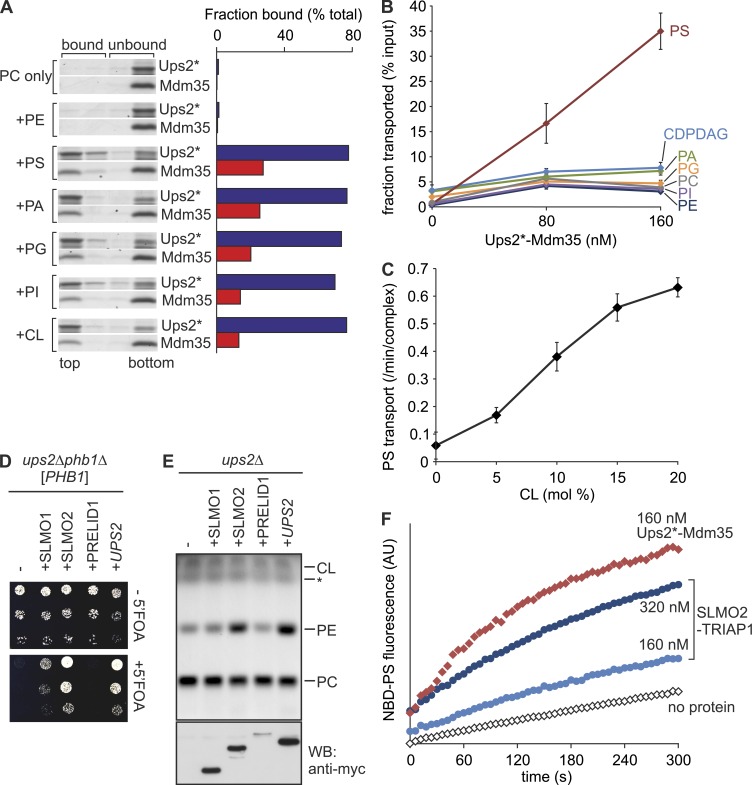
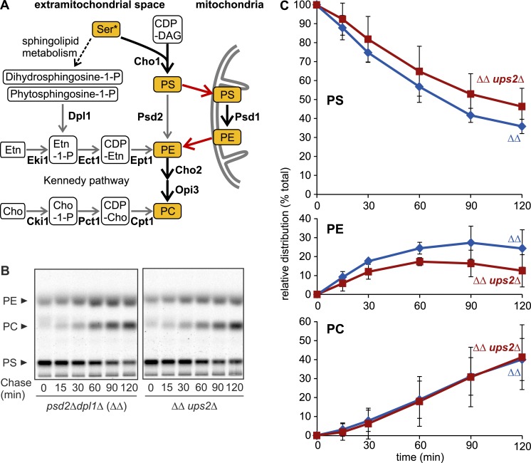
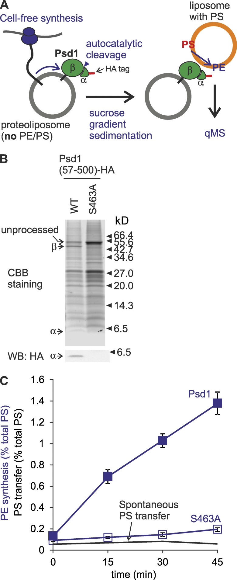
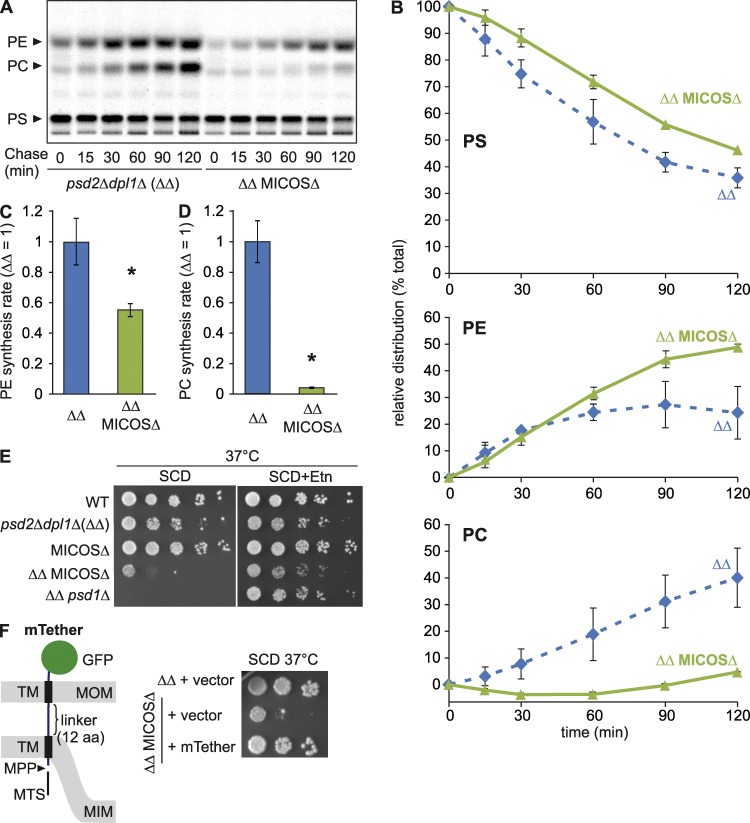
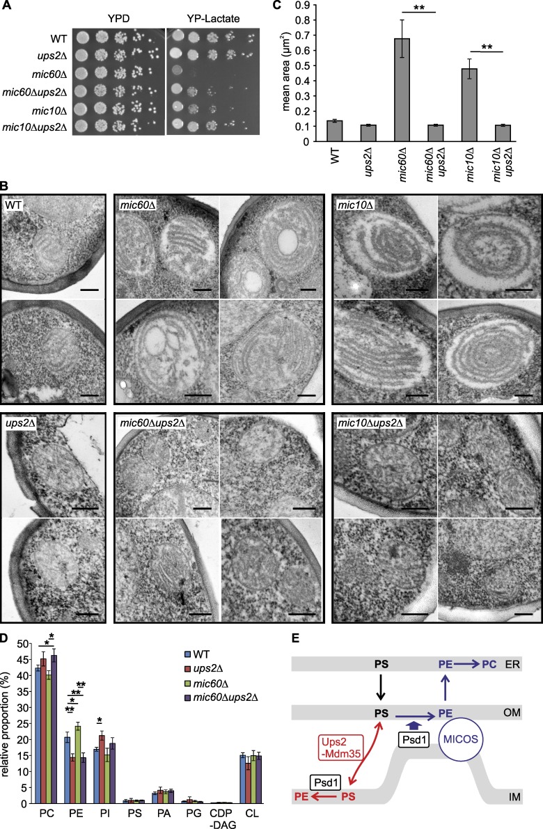
Mitochondria exert critical functions in cellular lipid metabolism and promote the synthesis of major constituents of cellular membranes, such as phosphatidylethanolamine (PE) and phosphatidylcholine. Here, we demonstrate that the phosphatidylserine decarboxylase Psd1, located in the inner mitochondrial membrane, promotes mitochondrial PE synthesis via two pathways. First, Ups2-Mdm35 complexes (SLMO2-TRIAP1 in humans) serve as phosphatidylserine (PS)-specific lipid transfer proteins in the mitochondrial intermembrane space, allowing formation of PE by Psd1 in the inner membrane. Second, Psd1 decarboxylates PS in the outer membrane in trans, independently of PS transfer by Ups2-Mdm35. This latter pathway requires close apposition between both mitochondrial membranes and the mitochondrial contact site and cristae organizing system (MICOS). In MICOS-deficient cells, limiting PS transfer by Ups2-Mdm35 and reducing mitochondrial PE accumulation preserves mitochondrial respiration and cristae formation. These results link mitochondrial PE metabolism to MICOS, combining functions in protein and lipid homeostasis to preserve mitochondrial structure and function.


Figures

Figure 1.
Figure 1.
Ups2–Mdm35 is a PS transfer protein complex. (A) Phospholipid binding. His-tagged Ups2C96S/C101S/C142S/C153S (Ups2*)–Mdm35 (5 µM) was incubated with liposomes composed of dioleoyl-PC (DOPC) and 20 mol% of the indicated dioleoyl-phospholipid (total lipid concentration 2 mM) for 10 min at 20°C. Upon flotation, four fractions were collected and analyzed by SDS-PAGE. Protein bands were analyzed densitometrically, and the sum of band intensities in all four lanes was set to 100%. The top two fractions represent bound material. (B) Phospholipid transfer. Ups2*–Mdm35 was incubated with donor liposomes (0.25 mM; DOPC/tetraoleoyl-CL/Lac-PE/17:0 PC/17:0 PE/DOPA/DOPS/DOPI/DOPG/DO-CDPDAG/DOPE/NBD-PE = 35/15/10/5/5/5/5/5/5/5/4.5/0.5 mol%, filled with 12.5% sucrose) and acceptor liposomes (1 mM, DOPC/DOPE/tetraoleoyl-CL/Lac-PE/rhodamine-PE = 50/24.95/15/10/0.05 mol%) for 5 min at 16°C. After isolation of acceptor liposomes by flotation, lipids were extracted and quantified by qMS. Error bars represent SEM. n = 4. (C) CL dependence of PS transfer activity by Ups2*–Mdm35. PS transfer was monitored by fluorescence dequenching (Fig. S1 C). Error bars represent SEM. n = 3. (D) Expression of human SLMO2 suppresses the lethality of ups2Δphb1Δ cells. Serial dilutions of ups2Δphb1Δ (PHB1) cells expressing C-terminally myc-tagged SLMO1, SLMO2, or PRELID1 were spotted on glucose-containing media with or without 5-fluoroorotic acid (5′FOA). (E) SLMO2 expression restores PE levels in Δups2 mitochondria. TLC of mitochondrial lipids. *, Unidentified lipid species. Same samples were subjected to SDS-PAGE and immunoblot analysis using anti-myc antibodies (WB). (F) SLMO2–TRIAP1 complex facilitates intermembrane transfer of NBD-PS. Transfer of NBD-PS was monitored by fluorescence dequenching. AU, arbitrary units.
Figure 2.
Figure 2.
PE biogenesis can proceed in the absence of Ups2. (A) PE/PC biogenesis in S. cerevisiae. Thick arrows represent the reactions proceeding in the experiments in B and in Fig. 4 A, where nonmitochondrial PE/PC synthesis was shut down by deletion of DPL1 and PSD2 and the lack of ethanolamine (Etn) or choline (Cho) in the medium. Red arrows represent lipid exchange. (B) Assessment of PE/PC synthesis in vivo. After pulse labeling with [14C]serine and further incubation in medium with unlabeled serine (chase), lipids were extracted and analyzed by TLC and autoradiography. ΔΔ, dpl1Δpsd2Δ. (C) Levels of PS, PE, and PC were quantified and are shown as a fraction of total lipids. Error bars represent SD. n = 3.
Figure 3.
Figure 3.
Psd1 is able to convert PS to PE at juxtaposed membranes in trans. (A) Monitoring of trans-decarboxylation by reconstituted Psd1. (B) Autocatalytic processing of reconstituted Psd1. The Psd1-containing fraction after flotation was subjected to SDS-PAGE and immunoblot analysis using HA-specific antibodies. CBB, Coomassie brilliant blue. (C) Psd1 decarboxylates PS to PE at juxtaposed membrane. S463A, Psd1S463A. Spontaneous PS transfer under identical conditions was plotted for comparison. PE produced or PS transferred at given time points are shown as percentage of total PS input. Error bars represent SEM. n = 3.
Figure 4.
Figure 4.
MICOS assists mitochondrial synthesis of PE and its conversion to PC in the ER. (A) Synthesis of PS, PE, and PC in cells labeled with [14C]serine. ΔΔ, dpl1Δpsd2Δ. (B) Relative distributions of radioactivity in PS, PE, or PC bands are represented as in Fig. 2 C. Error bars represent SD. n = 3. (C and D) Psd1-dependent formation of PE and PC from [14C]PS. Synthesis rates for PE (C) and PC (D) in cells lacking MICOS are shown. Synthesis rates were obtained by plotting kinetic changes in band intensities of the sum of PE and PC (C) or PC (D) and determining the slope of the fitted curve (between t = 15 min and t = 90 min). Mean values of ΔΔ were set to 1. Error bars represent SEM. n = 3. *, P < 0.05. (E) Loss of nonmitochondrial PE synthesis impairs growth of MICOSΔ cells. Cell growth was assessed at 37°C on glucose-containing SC medium (SCD) supplemented with ethanolamine (Etn; 10 mM) where indicated. WT, wild-type. (F) Expression of an artificial tether between two mitochondrial membranes (mTether) suppresses ethanolamine auxotrophy of cells lacking MICOS and nonmitochondrial PE synthesis. Left, schematic representation of mTether; right, cell growth on SCD at 37°C.
Figure 5.
Figure 5.
Deletion of UPS2 restores respiratory growth and mitochondrial architecture in cells lacking MICOS. (A) Cell growth on fermentable and nonfermentable carbon sources. Serial dilutions of the indicated strains were analyzed on YPD or YP-lactate plates at 30°C. WT, wild-type. (B) EM analysis of cells lacking Ups2, Mic60, and/or Mic10. Representative micrographs of cells grown on YP-lactate are shown. Bars, 200 nm. (C) Mean area of mitochondria in electron micrographs from indicated strains. Error bars represent SEM. n > 15. **, P < 0.01. (D) qMS of the mitochondrial phospholipidome in cells grown in lactate medium. Error bars represent SEM. n = 3. *, P < 0.05; **, P < 0.01. (E) Model for the roles of Ups2–Mdm35 and MICOS for Psd1-dependent PE synthesis.

References

    1. Alkhaja A.K., Jans D.C., Nikolov M., Vukotic M., Lytovchenko O., Ludewig F., Schliebs W., Riedel D., Urlaub H., Jakobs S., and Deckers M.. 2012. MINOS1 is a conserved component of mitofilin complexes and required for mitochondrial function and cristae organization. Mol. Biol. Cell. 23:247–257. 10.1091/mbc.E11-09-0774 - DOI - PMC - PubMed
    1. Birner R., Bürgermeister M., Schneiter R., and Daum G.. 2001. Roles of phosphatidylethanolamine and of its several biosynthetic pathways in Saccharomyces cerevisiae. Mol. Biol. Cell. 12:997–1007. 10.1091/mbc.12.4.997 - DOI - PMC - PubMed
    1. Choi J.Y., Wu W.I., and Voelker D.R.. 2005. Phosphatidylserine decarboxylases as genetic and biochemical tools for studying phospholipid traffic. Anal. Biochem. 347:165–175. 10.1016/j.ab.2005.03.017 - DOI - PubMed
    1. Connerth M., Tatsuta T., Haag M., Klecker T., Westermann B., and Langer T.. 2012. Intramitochondrial transport of phosphatidic acid in yeast by a lipid transfer protein. Science. 338:815–818. 10.1126/science.1225625 - DOI - PubMed
    1. Dee C.T., and Moffat K.G.. 2005. A novel family of mitochondrial proteins is represented by the Drosophila genes slmo, preli-like and real-time. Dev. Genes Evol. 215:248–254. 10.1007/s00427-005-0470-4 - DOI - PubMed

MeSH terms