. 2016 May 9:8:103.
doi: 10.3389/fnagi.2016.00103. eCollection 2016.

EPA/DHA and Vitamin A Supplementation Improves Spatial Memory and Alleviates the Age-related Decrease in Hippocampal RXRγ and Kinase Expression in Rats

Affiliations

EPA/DHA and Vitamin A Supplementation Improves Spatial Memory and Alleviates the Age-related Decrease in Hippocampal RXRγ and Kinase Expression in Rats

Anne Létondor et al. Front Aging Neurosci. .

Abstract

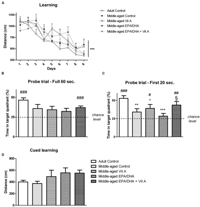
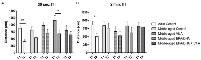
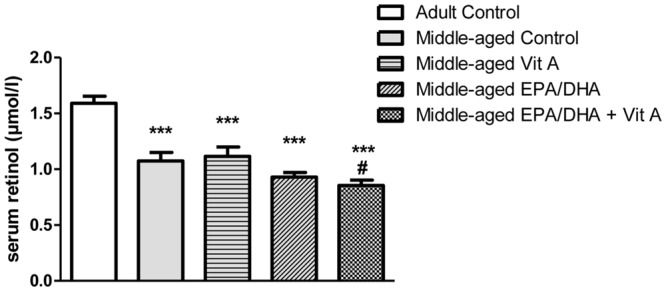
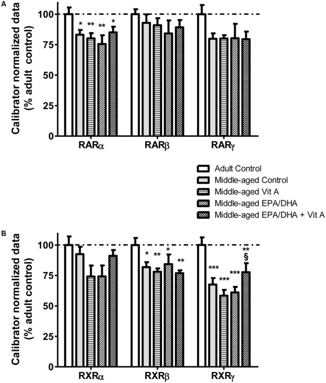
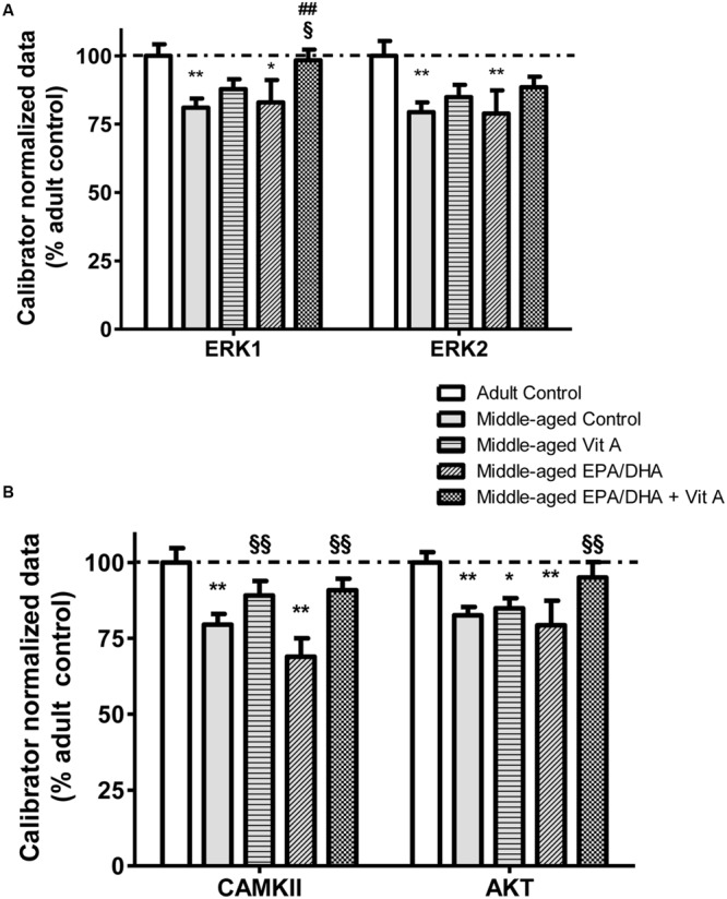
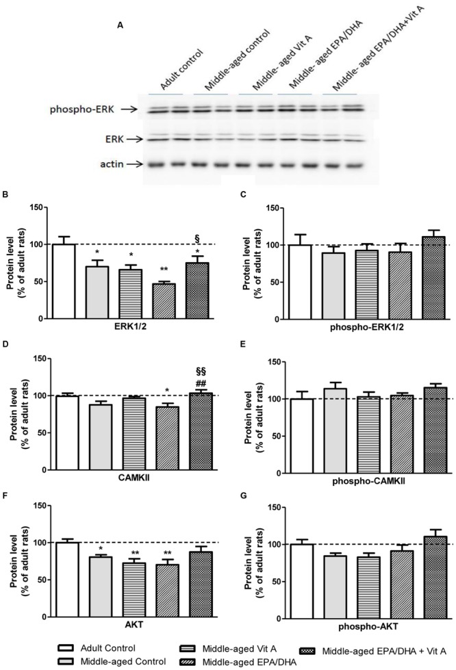
Studies suggest that eicosapentaenoic acid (EPA), docosahexaenoic acid (DHA), and vitamin A are critical to delay aged-related cognitive decline. These nutrients regulate gene expression in the brain by binding to nuclear receptors such as the retinoid X receptors (RXRs) and the retinoic acid receptors (RARs). Moreover, EPA/DHA and retinoids activate notably kinase signaling pathways such as AKT or MAPK, which includes ERK1/2. This suggests that these nutrients may modulate brain function in a similar way. Therefore, we investigated in middle-aged rats the behavioral and molecular effects of supplementations with EPA/DHA and vitamin A alone or combined. 18-month-old rats exhibited reference and working memory deficits in the Morris water maze, associated with a decrease in serum vitamin A and hippocampal EPA/DHA contents. RARα, RXRβ, and RXRγ mRNA expression and CAMKII, AKT, ERK1/2 expression were decreased in the hippocampus of middle-aged rats. A combined EPA/DHA and vitamin A supplementation had a beneficial additive effect on reference memory but not in working memory in middle-aged rats, associated with an alleviation of the age-related decrease in RXRγ, CAMKII, AKT, and ERK1 expression in the hippocampus. This study provides a new combined nutritional strategy to delay brain aging.

Keywords: hippocampus; kinases; n-3 long-chain PUFA; retinoid receptors; spatial memory; vitamin A.


Figures

FIGURE 1
FIGURE 1
Effect of age and diet (EPA/DHA, Vit A, or EPA/DHA + Vit A) on spatial learning, reference memory and cued learning in the Morris water maze. Figures show (A) the mean distance swum during acquisition to reach a submerged platform located at the same position over 9 days with four trials per day; (B,C) Percentage of time spent in the target quadrant with the platform removed in the probe test over (B) 60 s or (C) 20 s; (D) the mean distance swum during the cued learning. Values are mean ± SEM, n = 8–10 rats per group. Data were analyzed by (A) repeated measures ANOVA or (B–D) one-way ANOVA followed by the Fischer PLSD post hoc test. Signs indicate values different from (A) day 1 or (B,C) from Adult control: p < 0.05; ∗∗p < 0.01; ∗∗∗p < 0.001 and from middle-aged EPA/DHA: §p < 0.05; or compared with chance level by a Student’s one group t-test: #p < 0.05; ##p < 0.01; ###p < 0.001.
FIGURE 2
FIGURE 2
Effect of age and diet (EPA/DHA, Vit A, or EPA/DHA + Vit A) on spatial working memory (matching-to-place version) in the Morris water maze. Figures show (A) the mean distance swum to reach a submerged platform over 3 days in the first trial (T1) followed by a second trial (T2) with an inter-trial interval (ITI) of 30 s; (B) the mean distance swum to reach a submerged platform over 3 days in the T1 and T2 with an ITI of 2 min. The platform was moved to a different location each day. Values are mean ± SEM, n = 8–10 rats per group. Data were analyzed by Student’s paired t-test: different from T1: p < 0.05; ∗∗p < 0.01.
FIGURE 3
FIGURE 3
Effect of age and diet (EPA/DHA, Vit A, or EPA/DHA + Vit A) on serum retinol concentrations. Values are mean ± SEM, n = 8–10 rats per group. Data were analyzed by a one-way ANOVA followed by the Fischer PLSD post hoc test. Signs indicate values different from Adult control: ∗∗∗p < 0.001 and from middle-aged control: #p < 0.05.
FIGURE 4
FIGURE 4
Effect of age and diet (EPA/DHA, Vit A, or EPA/DHA + Vit A) on the mRNA expression of RARα, β, γ (A), and RXRα, β, γ (B). The mRNA levels are expressed as the percentage of Adult control mRNA expression of target/reference ratio normalized by the calibrator. Values are mean ± SEM, n = 8–10 rats per group. Data were analyzed by one-way ANOVAs followed by the Fischer PLSD post hoc test. Signs indicate values different from Adult control: p < 0.05; ∗∗p < 0.01; ∗∗∗p < 0.001 and from middle-aged Vit A and middle-aged EPA/DHA: §p < 0.05.
FIGURE 5
FIGURE 5
Effect of age and diet (EPA/DHA, Vit A, or EPA/DHA + Vit A) on the mRNA expression of ERK1, ERK2 (A) and CAMKII and AKT (B). The mRNA levels are expressed as the percentage of Adult control mRNA expression of target/reference ratio normalized by the calibrator. Values are mean ± SEM, n = 8–10 rats per group. Data were analyzed by one-way ANOVAs followed by the Fischer PLSD post hoc test. Signs indicate values different from Adult control: p < 0.05; ∗∗p < 0.01, from middle-aged control: ##p < 0.01 and from middle-aged EPA/DHA: §p < 0.05; §§p < 0.01.
FIGURE 6
FIGURE 6
Effect of age and diet (EPA/DHA, Vit A, or EPA/DHA + Vit A) on protein kinase expression in the hippocampus analyzed by Western blot. The upper panel (A) illustrates representative immunoblots probed for phospho-Erk1/2, Erk1/2 and actin. Hippocampal Erk1/2 (B), phospho-Erk1/2 (C), CAMKII (D), phospho-CAMKII (E), AKT (F), and phospho-AKT (G) protein levels were normalized against actin and expressed as a percentage of adult control expression of the same protein. Values are means ± SEM, n = 6–9 rats per group. Data were analyzed by one-way ANOVAs followed by the Fischer PLSD post hoc test. Signs indicate values different from Adult control: p < 0.05; ∗∗p < 0.01; from middle-aged control: ##p < 0.01 and from middle-aged EPA/DHA: §p < 0.05; §§p < 0.01.

Similar articles

Cited by

References

    1. Aid S., Bosetti F. (2007). Gene expression of cyclooxygenase-1 and Ca(2+)-independent phospholipase A(2) is altered in rat hippocampus during normal aging. Brain Res. Bull. 73 108–113. 10.1016/j.brainresbull.2007.02.015 - DOI - PMC - PubMed
    1. Akbar M., Calderon F., Wen Z., Kim H. Y. (2005). Docosahexaenoic acid: a positive modulator of Akt signaling in neuronal survival. Proc. Natl. Acad. Sci. U.S.A. 102 10858–10863. 10.1073/pnas.0502903102 - DOI - PMC - PubMed
    1. Al Tanoury Z., Piskunov A., Rochette-Egly C. (2013). Vitamin A and retinoid signaling: genomic and nongenomic effects. J. Lipid Res. 54 1761–1775. 10.1194/jlr.R030833 - DOI - PMC - PubMed
    1. Alfos S. (2014). “Fish oil supplementation prevents age-related memory decline: involvement of nuclear hormone receptors,” in Omega 3 Fatty Acids in Brain and Neurologic Health 1st Edn eds Watson R. R., De Meester F. (San Diego, CA: Elsevier; ) 147–161.
    1. Armbrecht H. J., Siddiqui A. M., Green M., Farr S. A., Kumar V. B., Banks W. A., et al. (2014). SAMP8 mice have altered hippocampal gene expression in long term potentiation, phosphatidylinositol signaling, and endocytosis pathways. Neurobiol. Aging 35 159–168. 10.1016/j.neurobiolaging.2013.07.018 - DOI - PMC - PubMed