

A Review of Sleep and Its Disorders in Patients with Parkinson's Disease in Relation to Various Brain Structures
- PMID: 27242523
- PMCID: PMC4876118
- DOI: 10.3389/fnagi.2016.00114
A Review of Sleep and Its Disorders in Patients with Parkinson's Disease in Relation to Various Brain Structures
Abstract
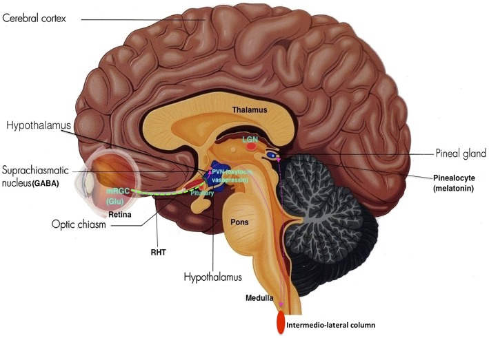
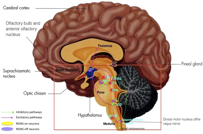
Sleep is an indispensable normal physiology of the human body fundamental for healthy functioning. It has been observed that Parkinson's disease (PD) not only exhibits motor symptoms, but also non-motor symptoms such as metabolic irregularities, altered olfaction, cardiovascular dysfunction, gastrointestinal complications and especially sleep disorders which is the focus of this review. A good understanding and knowledge of the different brain structures involved and how they function in the development of sleep disorders should be well comprehended in order to treat and alleviate these symptoms and enhance quality of life for PD patients. Therefore it is vital that the normal functioning of the body in relation to sleep is well understood before proceeding on to the pathophysiology of PD correlating to its symptoms. Suitable treatment can then be administered toward enhancing the quality of life of these patients, perhaps even discovering the cause for this disease.
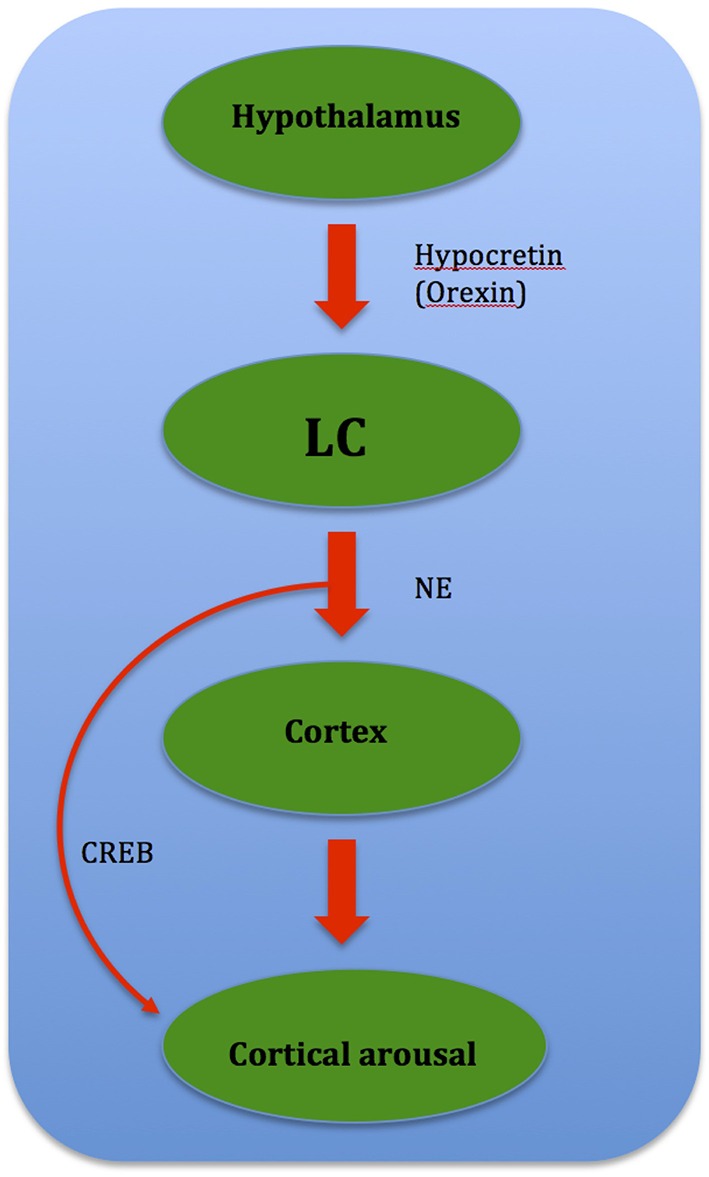
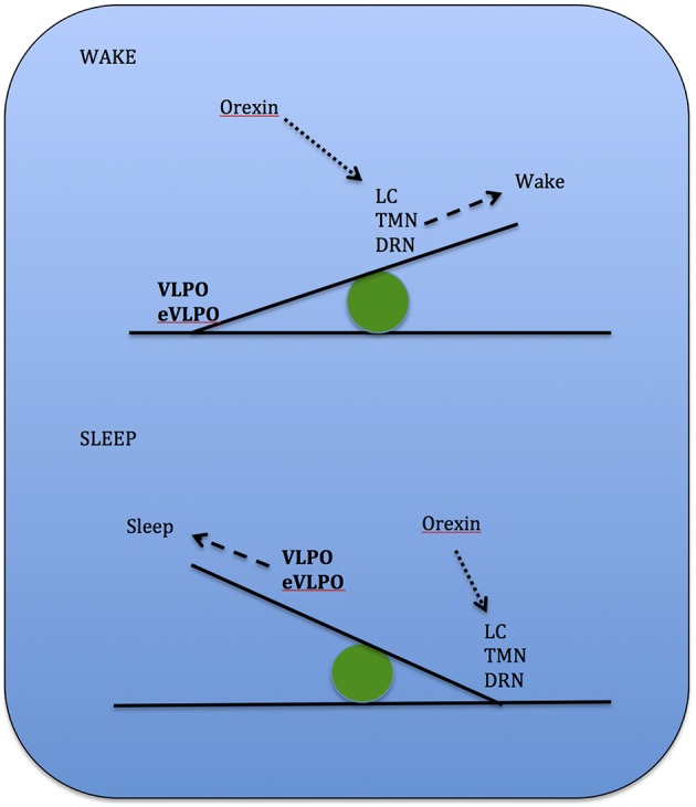
Keywords: Parkinson's disease (PD); REMS behavior disorder (RBD); dopamine; hypocretin; non-rapid eye movement sleep (NREMS); rapid eye movement sleep (REMS); sleep; suprachiasmatic nucleus (SCN).
Figures
Similar articles
-
[Syndrome of rapid eye movement sleep behavior disorder and nocturia in Parkinson's disease].Zh Nevrol Psikhiatr Im S S Korsakova. 2017;117(9):15-20. doi: 10.17116/jnevro20171179115-20. Zh Nevrol Psikhiatr Im S S Korsakova. 2017. PMID: 29053115 Russian.
-
Rapid Eye Movement Sleep Behavior Disorder in Parkinson's Disease: A Preliminary Study.J Mov Disord. 2016 May;9(2):114-9. doi: 10.14802/jmd.15039. Epub 2016 Mar 2. J Mov Disord. 2016. PMID: 26936443 Free PMC article.
-
Frequency of REM sleep behavior disorders in patients with Parkinson's disease.Vojnosanit Pregl. 2015 May;72(5):442-6. doi: 10.2298/vsp130501006j. Vojnosanit Pregl. 2015. PMID: 26165053
-
[Selective stimulations and lesions of the rat brain nuclei as the models for research of the human sleep pathology mechanisms].Glas Srp Akad Nauka Med. 2011;(51):85-97. Glas Srp Akad Nauka Med. 2011. PMID: 22165729 Review. Serbian.
-
Clinical Significance of REM Sleep Behavior Disorders and Other Non-motor Symptoms of Parkinsonism.Neurosci Bull. 2017 Oct;33(5):576-584. doi: 10.1007/s12264-017-0164-8. Epub 2017 Aug 3. Neurosci Bull. 2017. PMID: 28770440 Free PMC article. Review.
Cited by
-
Poor nighttime sleep is positively associated with dyskinesia in Parkinson's disease patients.Parkinsonism Relat Disord. 2018 Mar;48:68-73. doi: 10.1016/j.parkreldis.2017.12.022. Epub 2017 Dec 21. Parkinsonism Relat Disord. 2018. PMID: 29305084 Free PMC article.
-
Circadian clock disruption promotes the degeneration of dopaminergic neurons in male Drosophila.Nat Commun. 2023 Sep 22;14(1):5908. doi: 10.1038/s41467-023-41540-y. Nat Commun. 2023. PMID: 37737209 Free PMC article.
-
Disrupted sleep-wake regulation in the MCI-Park mouse model of Parkinson's disease.NPJ Parkinsons Dis. 2024 Mar 11;10(1):54. doi: 10.1038/s41531-024-00670-w. NPJ Parkinsons Dis. 2024. PMID: 38467673 Free PMC article.
-
Acute sleep deprivation in mice generates protein pathology consistent with neurodegenerative diseases.Front Neurosci. 2024 Jul 24;18:1436966. doi: 10.3389/fnins.2024.1436966. eCollection 2024. Front Neurosci. 2024. PMID: 39114483 Free PMC article.
-
Rotigotine transdermal patch and sleep in Parkinson's disease: where are we now?NPJ Parkinsons Dis. 2017 Sep 5;3:28. doi: 10.1038/s41531-017-0030-4. eCollection 2017. NPJ Parkinsons Dis. 2017. PMID: 28890931 Free PMC article. Review.
References
-
- Albanese A., Altavista M., Rossi P. (1985). Organization of central nervous system dopaminergic pathways. J. Neural Transm. Suppl. 22, 3–17. - PubMed
Publication types
LinkOut - more resources
Full Text Sources
Other Literature Sources